
图1")
图2")
图3")
图4")
图5")


| 图片: | |
|---|---|
| 名称: | |
| 描述: | |
- B22Breast papillary lesion cqz (1)
图1") 图1
图1图2") 图2
图2图3") 图3
图3图4") 图4
图4图5") 图5
图5
| 姓 名: | ××× | 性别: | f | 年龄: | 52 |
| 标本名称: | Breast segmental mastectomy | ||||
| 简要病史: | Breast lesion | ||||
| 肉眼检查: | |||||
failed to poste the photos and try again.
Your diagnosis
Differential diagnoses
What immunostains will be useful?
标签:
-
本帖最后由 于 2009-02-17 09:36:00 编辑
×参考诊断
DCIS involving the papilloma(DCIS累犯乳头状瘤)
-
本帖最后由 于 2008-10-29 18:16:00 编辑
使用ER帮助诊断乳头状病变,来自陈国璋教授的讲课资料。
Papilloma or Papillary DCIS / intracystic papillary carcinoma?
--One of the most difficult diagnostic problems
--Solving problem by immunohistochemistry:
Abundant myoepithelial cells in the cores of papillae in papilloma, but not in the papillae of papillary carcinoma [Note: Myoepithelium is absent around the cyst in intracystic papillary carcinoma]
CK5 (preserved in papilloma)
ER (uniform strong staining favors carcinoma)

华夏病理/粉蓝医疗
为基层医院病理科提供全面解决方案,
努力让人人享有便捷准确可靠的病理诊断服务。
-
本帖最后由 于 2008-10-30 23:35:00 编辑
To Dr. Abin:
Agree that myoepithelial markers are useful for the differential dx of papillary lesions.
I am not sure the ER, as I mentioned that I have no experience about ER in papillary lesion. My impression is that epithelial cells in most papilloma cases are also positive for ER. Could you let me know the original study (not text book) papers or study results. Hope to learn the differences in details about the positive rate of ER ,and extention and intensity of the stains among papilloma, atypical papilloma, and papillary DCIS in these studies.
Thanks
cqz
WHO: Introductal papillary neoplasms: it mentioned the following 5 types
Central papilloma
Peripheral papilloma
Atypical papilloma
Intraductal papillary carcinoma
Intracystic papillary carcinoma
I copied the original sentence from WHO book about the definition of intraductal papillary carcinoma:
Intraductal papillary carcinoma's diagnosis requires that 90% ir more of the papillary processes are totally devoid of a myoepithelial cell layer regardless of presence or absence of notavle epithelial proliferation, and/or that any of the recognized patterns of low grade DCIS occupies 90% or more of the lesion.
So the lesions between atypical papilloma and intraductal papillary carcinoma will be called as DCIS involving or arising from papilloma, even though it is not mentioned in WHO book. But it has be used for a long time in the literature and in the clinical diagnosis.
So DCIS involving papilloma and intraductal papillary ca are the same disease process with the different degree. The botoom line is that they are kinds of of DCIS with the same clinical managment.
Hope it can help,
To Abin and Lili:
ER for differential dx of papillary lesion.
I asked one of our previous GYN/Breast fellow who did a lot of research in papillary lesions and works as a breast pathologist in another institute now. I copied her response below.
I am unaware of any significant literature on using ER in the differential of papilloma, atypical papilloma, and pap DCIS. I know some people use neuroendocrine markers to help in the diff of atypical and pap, but the yield is low on that. THe best IHC markers to help in the differential, at least according to Schnitt and Collins, are myoep markers, since papillomas should have a sprinkling of myoeps throughout, atypical papillomas have foci of absent myoep markers but still have myoeps lining the fibrovascular cores, and pap DCIS should be COMPLETELY devoid of myoep markers (except at the periphery of course...except for encapsulated papillary carcinomas!)...hope that makes sense.
Above is her answer.
Any way it makes no sense for me to use ER in the differentiation. There are more ductal epithelial proliferation in DCIS or papillary ca than papilloma. ER is positive in ductal epithelial and negative for myoepthelial cells. This is why you may feel stong and diffuse stain in DCIS in papilloma or papillary ca. I do not think ER is usful for differential dx in papillary lesions untill you show me the original reference papers with solid evidence.
Just for your reference.
-
本帖最后由 于 2008-10-30 23:49:00 编辑
回赵老师:请原谅我不求甚解,我是学习了陈国璋教授的讲课资料后,在实际工作中验证并接受了这个观点。不仅是乳头状病变,ER对UDH和低级别DCIS的鉴别也有帮助。我只有陈国璋教授的资料,其他的数据和文献就无法提供啦。
sorry again
Usual ductal hyperplasia: Key features
“Mixed and disorderly”
Interspersed spaces vary in size and shape; spaces tend to be peripherally located
Cells often have indistinct cell borders, and are unevenly spaced
Nuclei are often oval, with grooves and pseudoinclusions
Nuclei show streaming
May have some admixed apocrine cells
Cytokeratin profile: mixed (CK5+, CAM5.2+)
ER: heterogeneous
Low-grade DCIS: Key features
“Uniform”
Interspersed spaces, if present, are round and rigid
Cells often have distinct cell borders, and are evenly spaced
Nuclei are often round, with hyperchromasia
Pagetoid spread (lifting up residual luminal cells), if present, favors diagnosis of DCIS
Cytokeratin profile: one type of cells -- glandular cells (CK5-, CAM5.2+)
ER: usually uniform strong
华夏病理/粉蓝医疗
为基层医院病理科提供全面解决方案,
努力让人人享有便捷准确可靠的病理诊断服务。
Papilloma-atypical papilloma-papillary ca
UDH-ADH-DCIS
No solid research data support that IHC stains are usful for the differential dx. Several years ago there were some studies indicating the IHC including high molecular cytokeratin may be useful. In term of my knowledge few people use IHC for differential dx of these lesions in the US now. Of cause you can continue to use them if you feel they have some usage in your clinical practice. You can summary your data for a publication if the results are good.
Thanks
-
本帖最后由 于 2008-11-04 20:24:00 编辑
 图1
图1 图2
图2
I put another breast lesion here.
50 year women with breast mass
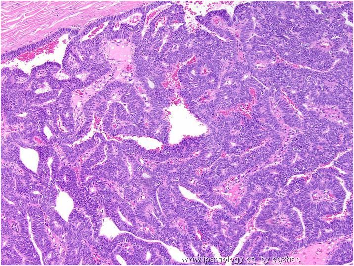
Fig 1 is a low power view of the lesion, where the overlying skin adjacent to the nipple is seen overlying a well-circumscribed mass.
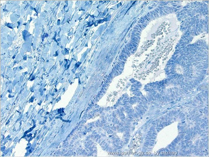
Fig 2 mid power
Fig 3 Hihg power
What is your diagnosis or differential dx?
Do you need IHC?
abin译:第33、34和36楼为另一例乳腺病变。患者为50岁女性,发现乳腺肿块。
图1为病变低倍观,示界清肿块,上方为乳头附近的皮肤。图2为中倍,图3为高倍。
您的诊断或鉴别诊断?是否需要免疫组化?


