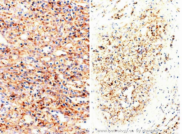
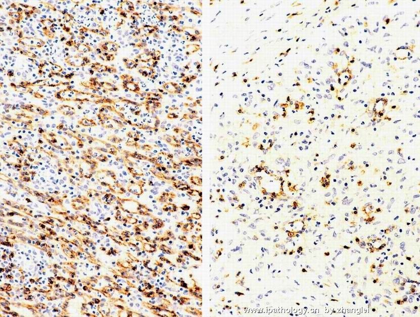
Sclerosing Angiomatoid Nodular Transformation (SANT): Report of 25 Cases of a Distinctive Benign Splenic Lesion.
American Journal of Surgical Pathology. 28(10):1268-1279, October 2004.
Martel, Maritza MD *; Cheuk, Wah MD +; Lombardi, Luciano MD *; Lifschitz-Mercer, Beatriz MD ++; Chan, John K. C MD +; Rosai, Juan MD *
Abstract:
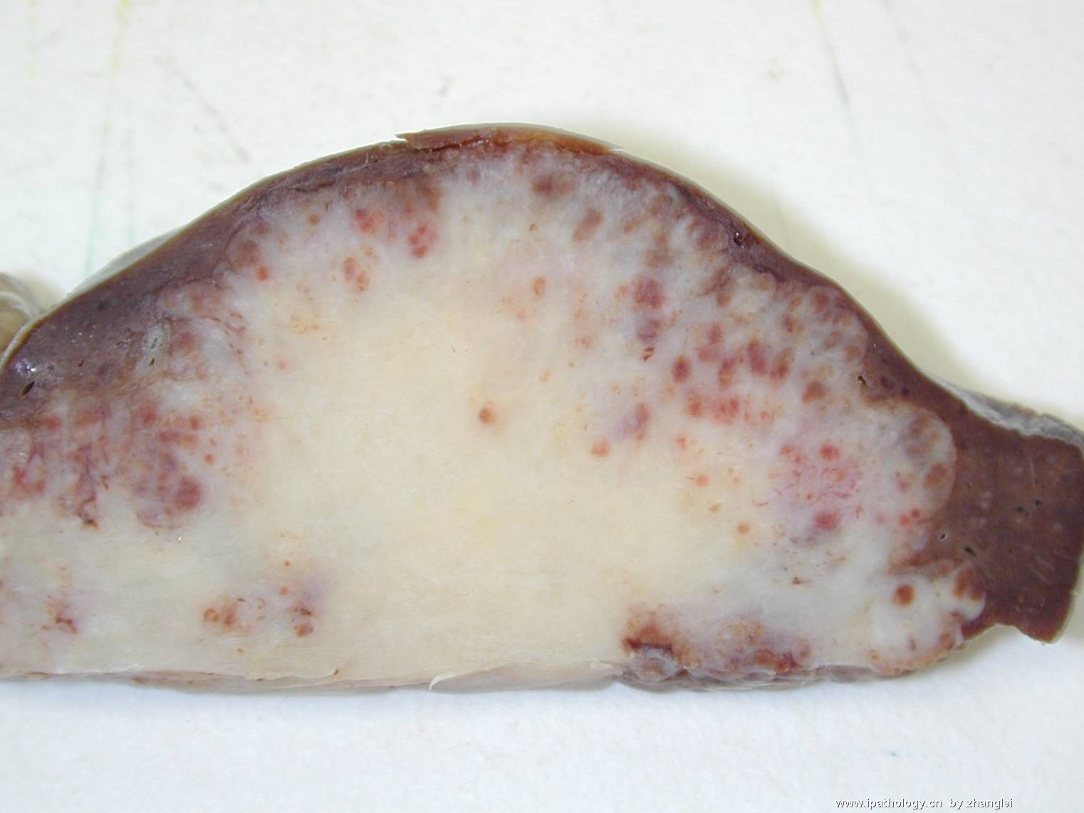
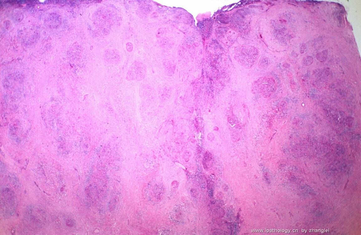
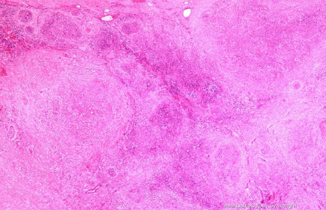
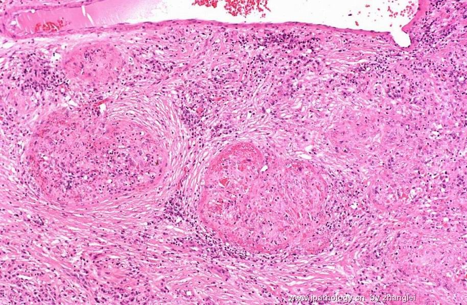
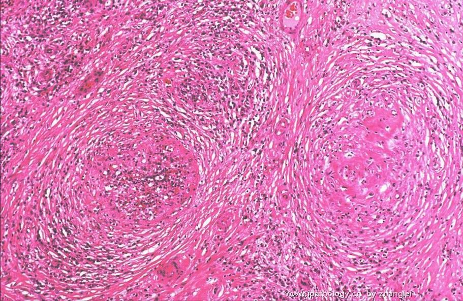
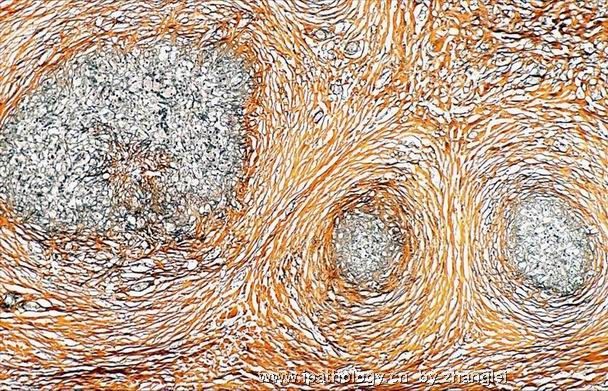
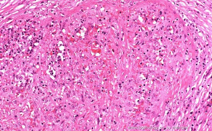
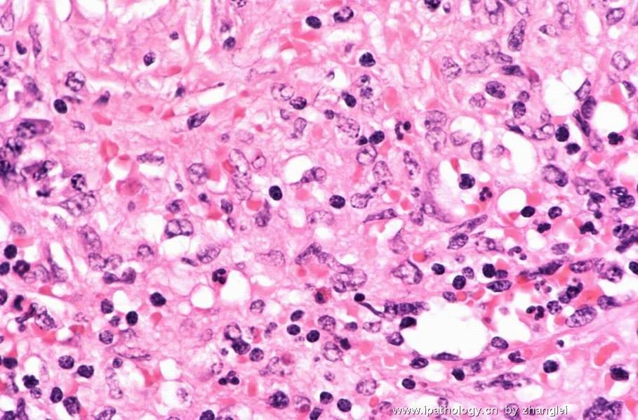
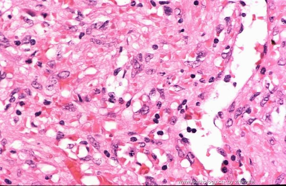
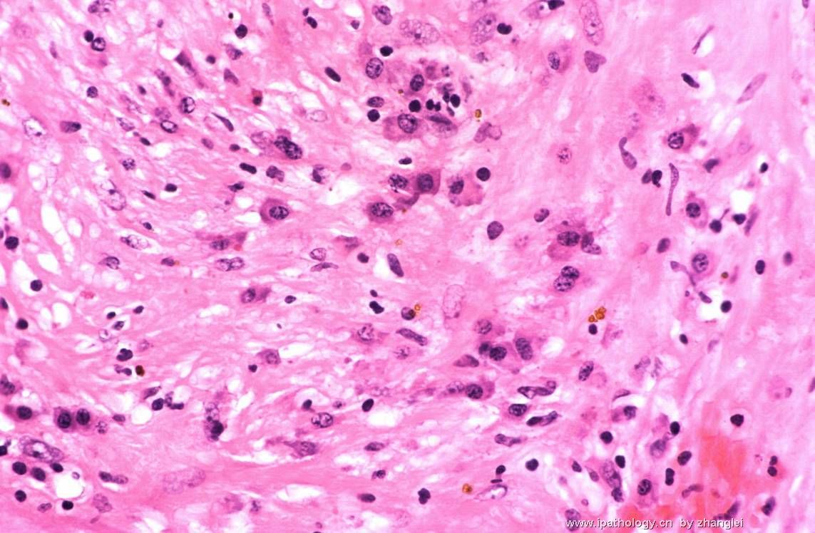
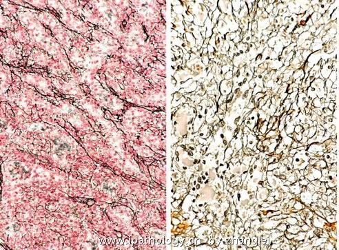
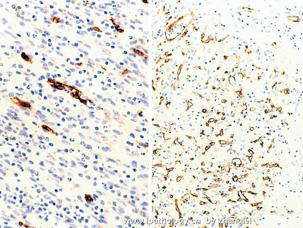
Twenty-five cases of a morphologically distinctive vascular lesion of the spleen are described. The patients were 17 women and 8 men, ranging in age from 22 to 74 years (mean, 48.4 years; median, 56 years). The most common presentations were incidental finding of an asymptomatic splenic mass (13 patients), abdominal pain or discomfort (6 patients), and splenomegaly (4 patients). None of the patients had evidence of recurrent disease after splenectomy. The splenic lesion was solitary, measuring 3 to 17 cm, and sharply demarcated from the surrounding parenchyma. The cut surface revealed a mass of coalescing red-brown nodules embedded in a dense fibrous stroma. All cases showed a remarkably consistent multinodular appearance at low-power examination. The individual nodules had an angiomatoid appearance, in the sense that they were composed of slit-like, round or irregular-shaped vascular spaces lined by plump endothelial cells and interspersed by a population of spindly or ovoid cells. Some of the nodules (particularly the smaller ones) were surrounded by concentric rings of collagen fibers. Numerous red blood cells were present, as well as scattered inflammatory cells. Nuclear atypia was minimal, mitotic figures were extremely rare, and necrosis was consistently absent. The internodular stroma consisted of variably myxoid to dense fibrous tissue with scattered plump myofibroblasts, plasma cells, lymphocytes, and siderophages. Immunostaining revealed 3 distinct types of vessels in the angiomatoid nodules: CD34+/CD8-/CD31+ capillaries, CD34-/CD8+/CD31+ sinusoids, and CD34-/CD8-/CD31+ small veins, recapitulating the composition of the normal splenic red pulp. These features are therefore different from those of littoral cell angioma, conventional hemangioma, and hemangioendothelioma of the spleen. We interpret these angiomatoid nodules as altered red pulp tissue that had been entrapped by a nonneoplastic stromal proliferative process. The characteristic morphologic appearance, immunophenotype, and benign clinical course suggest that this is a distinctive nonneoplastic vascular lesion of the spleen that we propose to designate as sclerosing angiomatoid nodular transformation (SANT).
(C) 2004 Lippincott Williams & Wilkins, Inc.
American Journal of Surgical Pathology. 21(7):827-835, July 1997.
Arber, Daniel A. M.D.; Strickler, John G. M.D.; Chen, Yuan-Yuan B.S.; Weiss, Lawrence M. M.D.
Abstract:
Vascular tumors of the spleen include several different entities, some of which are unique to that organ. Twenty-two such proliferations were studied, including 10 hemangiomas, six littoral cell angiomas, four angiosarcomas, and two hamartomas. The hemangiomas included seven with localized tumors and three with diffuse angiomatosis of the spleen. All cases were studied by paraffin section immunohistochemistry with a large panel of antibodies. In addition, all cases were studied for the presence of the Kaposi's sarcoma-associated herpesvirus (KSHV) using the polymerase chain reaction. The morphologic findings were similar to those previously reported. All proliferations were vimentin positive, and one angiosarcoma was focally keratin positive. All cases reacted for CD31, whereas 20 of 22 were positive for von Willebrand's factor and 19 of 22 were positive for Ulex europeaus. CD34 expression in lining cells was identified in 10 of 10 hemangiomas, two of four angiosarcomas, and one of two hamartomas, whereas all six cases of littoral cell angioma were negative. CD68 was expressed in all cases of littoral cell angioma but was also positive in all three diffuse hemangiomas, two of seven localized hemangiomas, and two of four angiosarcomas. CD21 expression was restricted to the lining cells of littoral cell angioma, and CD8 expression was only identified in two of two hamartomas and two of four angiosarcomas. KSHV was not detected in any of the cases. These findings suggest that there are distinct immunophenotypic as well as morphologic features of splenic vascular tumors. Littoral cell angiomas have a characteristic CD34-/CD68+/CD21+/CD8- immunophenotype and hamartomas have a characteristic CD68-/CD21-/CD8+ phenotype. The frequent CD68 expression in diffuse hemangioma suggests an immunophenotypic difference from localized hemangioma of the spleen.
