| 图片: | |
|---|---|
| 名称: | |
| 描述: | |
- 胰尾部囊肿(转帖)
| 以下是引用HHX 在2007-2-12 12:54:00的发言: 很想看看这个病例的肾和肝的影像学图片,不知能否提供?还有病史? |
Clinical History:
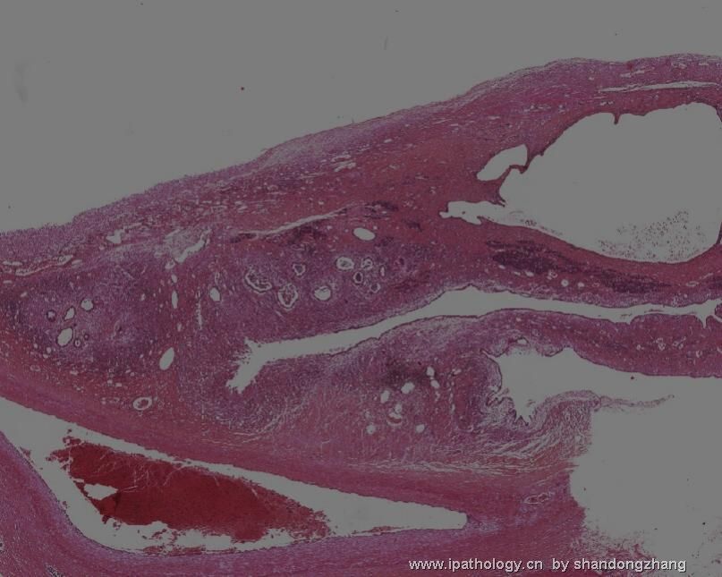
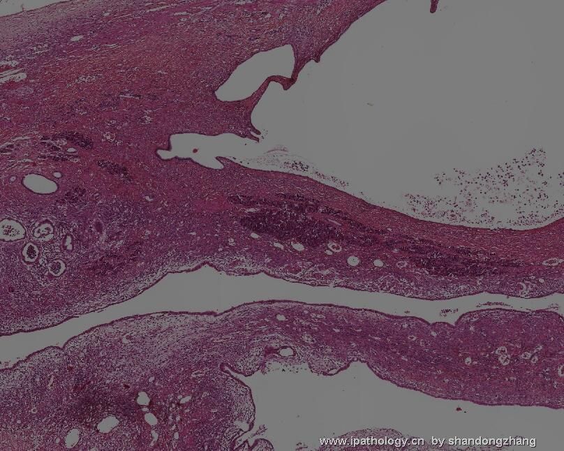
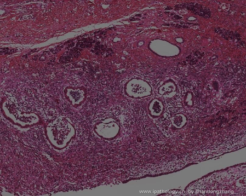
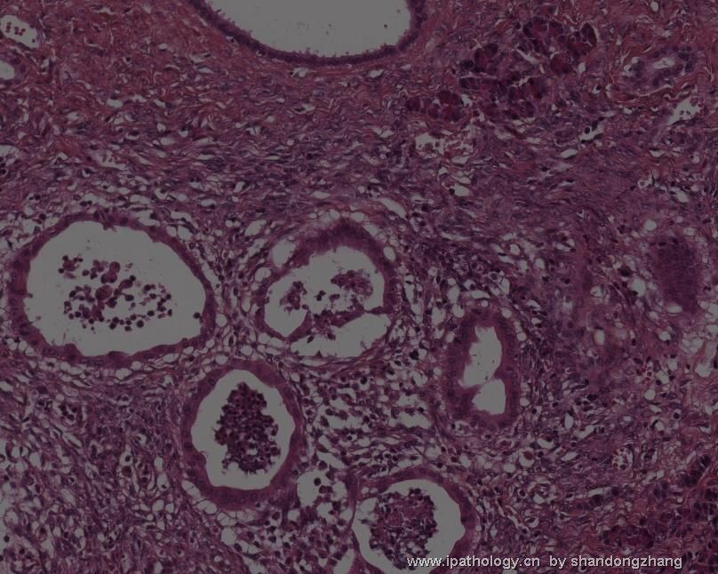
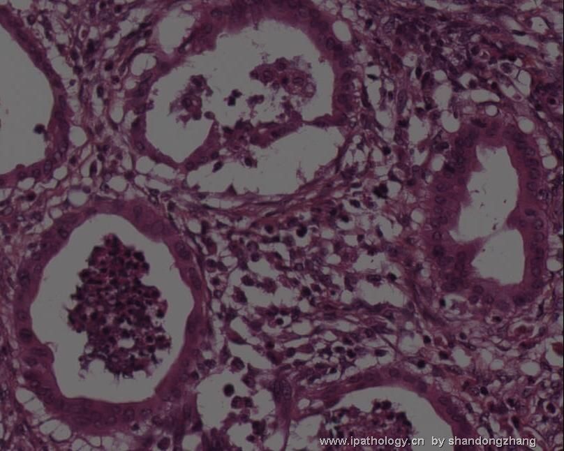
A 22-year-old female with no medical history was admitted to our hospital for further examination of anemia. She had no evidence of gastrointestinal bleeding such as hematemesis. On admission, she was slightly pale and an abdominal mass was palpable. Laboratory examination confirmed the anemia (haemoglobin 7.4g/dl, hematocrit 25;2% and MCV 85 fl). Upper gastrointestinal endoscopy revealed nodular oesophageal and gastric varices with red spots suggesting stigmata of bleeding. On abdominal CT, the tail of the pancreas revealed a 10cm unilocular cystic lesion with irregular walls and septations. Mild splenomegaly, obstruction of splenic vein and gastrooesophageal varices were also observed. A clinical diagnosis of pancreatic cystadenocarcinoma was proposed. The patient underwent a distal pancreatectomy. Macroscopically, the tail of pancreas was occupied by a 10cm unilocular cyst showing numerous septa and filled with serosanguineous fluid. The inner lining of the cystic space did not show nodular masses. The selected slide concerns the cyst wall.

