| 图片: | |
|---|---|
| 名称: | |
| 描述: | |
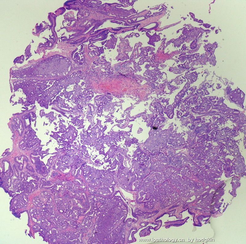
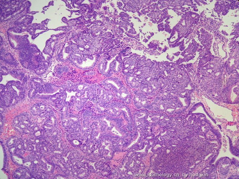
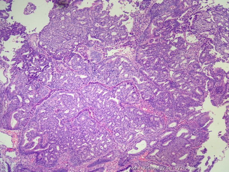
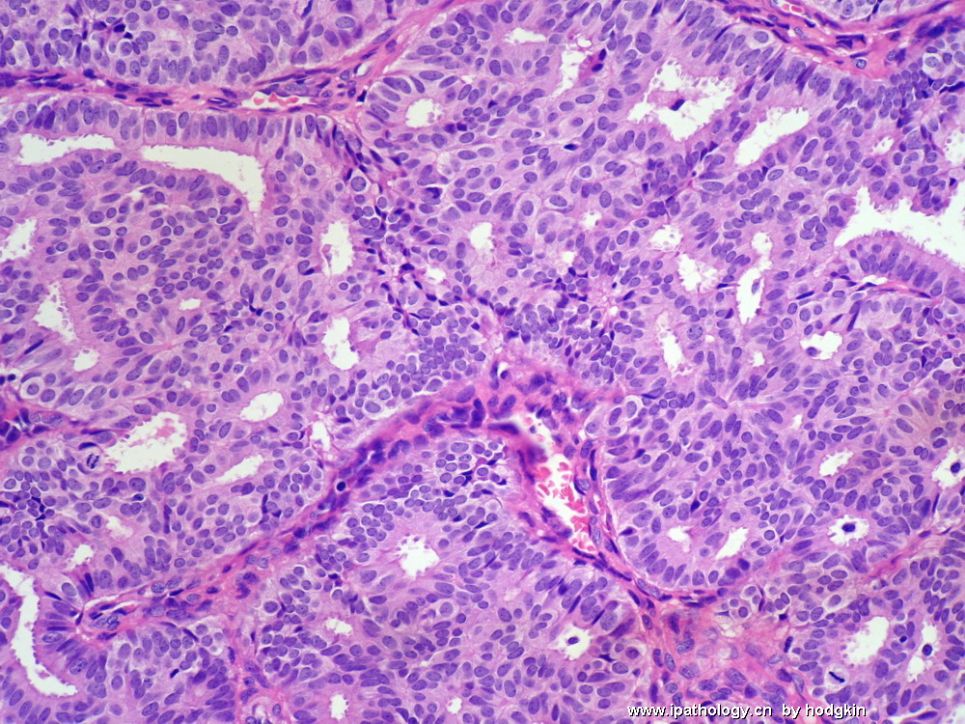
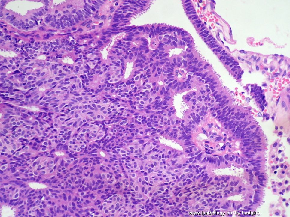
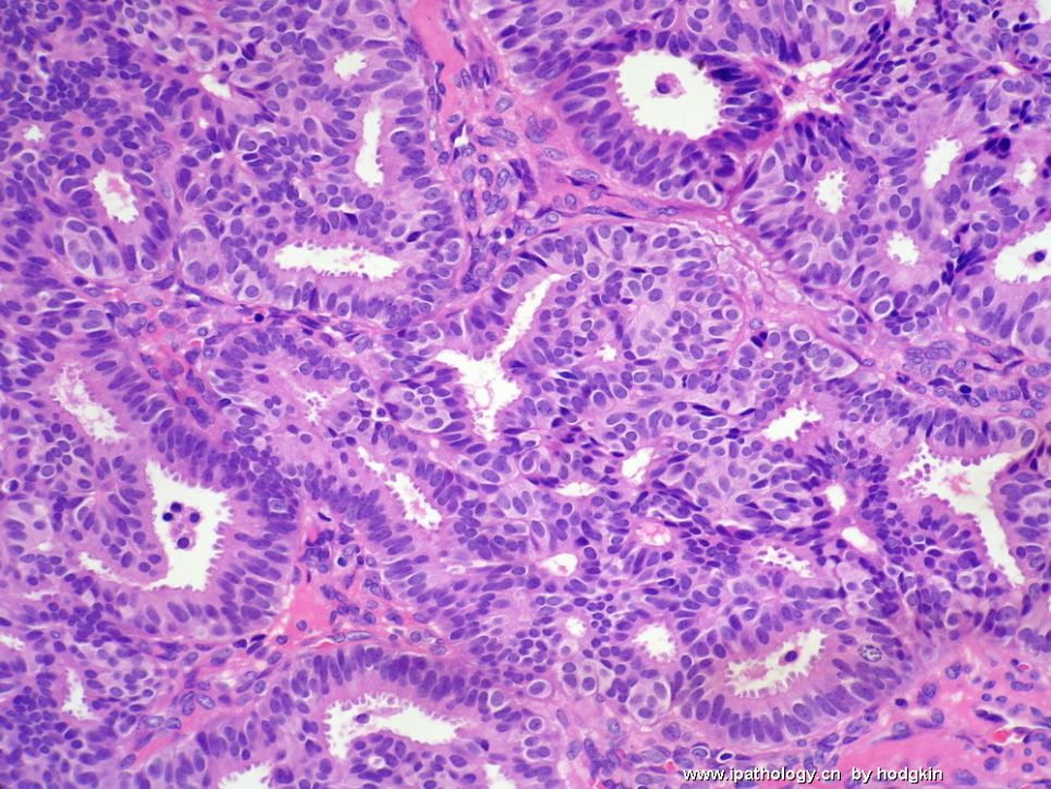
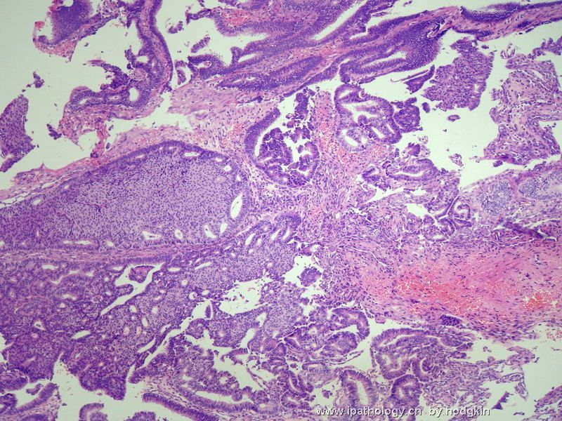
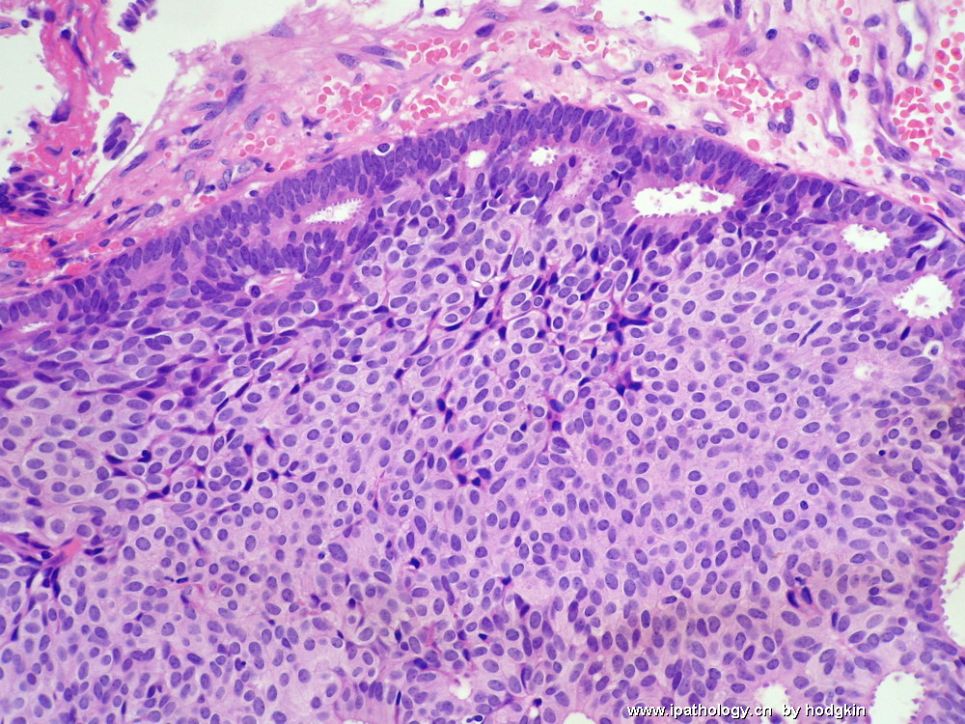
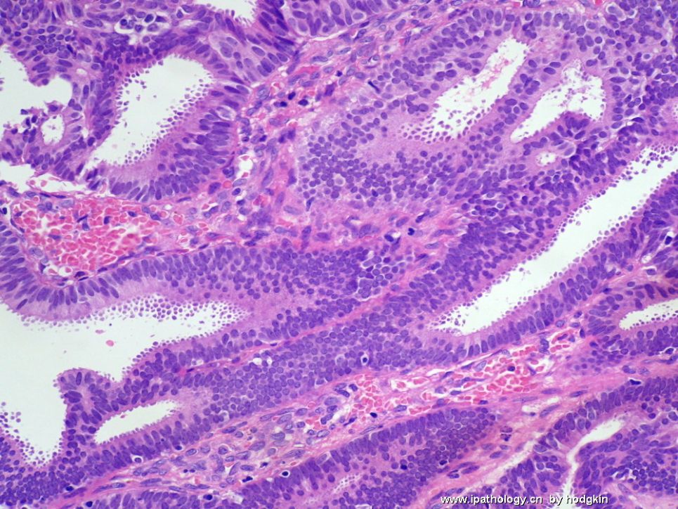
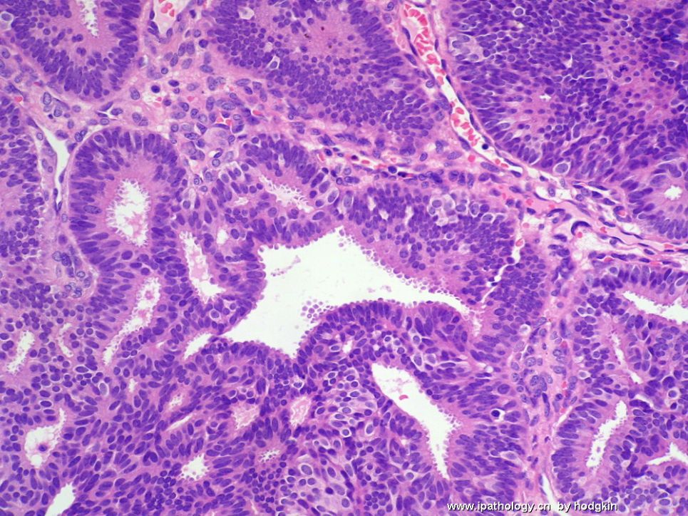
- 57岁女性,乳腺肿物。
-
stevenshen 离线
- 帖子:343
- 粉蓝豆:2
- 经验:343
- 注册时间:2008-06-03
- 加关注 | 发消息
Hi hodgkin, thanks for hosting the lecture for me. I am not an expert on breast pathology, but would like to get better at it. I have made some comment before on this case before. If you don't mind, I would make additional observations. I enjoyed your high quality pictures. I hope that you will share with us your final answer...
Unless you worry about invasive carcinoma elsewhere, I don't think the immunostains you provided will change the diagnosis of either DCIS and ADH. I still think it is a low grade DCIS, particularly the 2 additional frozen pictures. I knew that frozen section of breast tissue is commonly performed in China, which is very different from the practice in USA. We almost never do frozen section on breast cancer...just imaging all the difficult situations - UDH vs DCIS vs ADH vs LCIS, sclersoing adenosis with involvement of LCIS or DCIS vs. invasive carcinoma, invasive tubular or lobular carcinomas...Doing a frozen section on these small breast lesions is really like "walk on the ice". You are luck if you don't fall. Thanks!
-
I totally agree with Dr. Shen. Low grade DCIS. None of the hospitals i worked in US does breast frozen sections routinely. We do touch prep sometimes on a mass and gross assessement of margins. Of course i do touch prep and frozen sections of sentinel lymph nodes.
I am not convinced that DCIS is present. The pictures 6, 7 in the first group of photos and the first and secon in the fozen section show focal monotonous and may be diagnosed as atypical ductal hyperplasia. It is better to see the photo one and two in f frozen sections in high power and also know the size of uniform area.
Based on the photos, my impression:
ADH
UDH
CCC
IHC cannot help you.
-
stevenshen 离线
- 帖子:343
- 粉蓝豆:2
- 经验:343
- 注册时间:2008-06-03
- 加关注 | 发消息
-
This is a difficult case to interpret. The reason is because there are a lots processes going on at the same time. It is complex proliferative lesion including usual ductal hyperplasia, columnar cell changes and hyperplasia. I agree with many people also that there are component of atypical ductal hyperplasia (ADH) involving almost the entire lesion (1 cm). The debate here is probably ADH or ductal carcinoma in situ (DCIS). I would characterize this as low grade DCIS, solid and cribriform growth patterns. From the pictures, I see no convincing evidence of invasion. I look forward to hear the final diagnosis. Very nice images. Thanks!
| 以下是引用stevenshen在2008-9-22 7:20:00的发言: This is a difficult case to interpret. The reason is because there are a lots processes going on at the same time. It is complex proliferative lesion including usual ductal hyperplasia, columnar cell changes and hyperplasia. I agree with many people also that there are component of atypical ductal hyperplasia (ADH) involving almost the entire lesion (1 cm). The debate here is probably ADH or ductal carcinoma in situ (DCIS). I would characterize this as low grade DCIS, solid and cribriform growth patterns. From the pictures, I see no convincing evidence of invasion. I look forward to hear the final diagnosis. Very nice images. Thanks! |

