| 图片: | |
|---|---|
| 名称: | |
| 描述: | |
- 子宫内膜
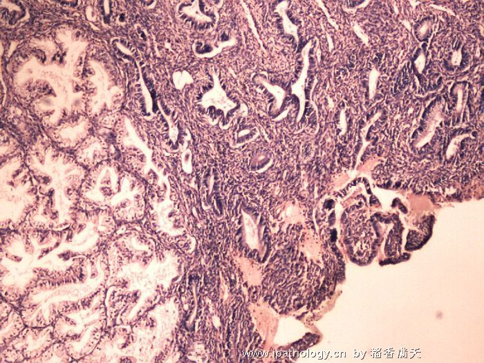
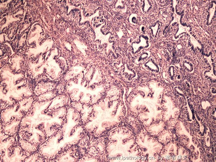
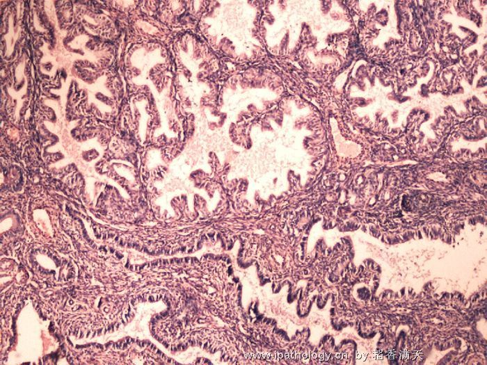
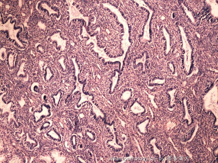
| 以下是引用cqzhao在2008-10-5 9:00:00的发言: Cannot see the lesion in lower power. feel it may be a benign endometrial polyp with focal secretory change- functional endometrial polyp. I once asked Dr. Kurman the question. If endometrial polys show complex hyperplasia without atypia, you do not need to mention the complex hyperplasia. The terminology complex hyperplasia may scare the physicians or patients. Follow-up data show CH without atypia in polys with no affect to the prognosis. Just report endometrial polyp. However you have to report it if the nuclear atypia is present. Do not matter it is simple hyperplasia or no hyperplasia. Thanks for your analysis! |

朱正龙
-
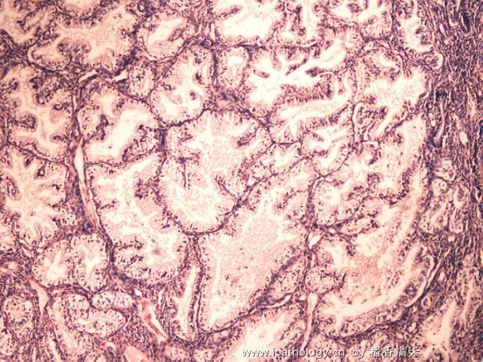
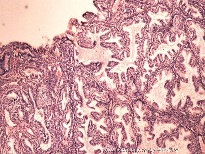
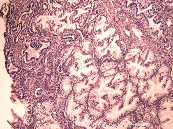
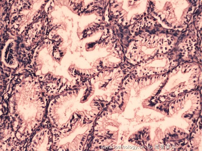
Cannot see the lesion in lower power. feel it may be a benign endometrial polyp with focal secretory change- functional endometrial polyp. I once asked Dr. Kurman the question. If endometrial polys show complex hyperplasia without atypia, you do not need to mention the complex hyperplasia. The terminology complex hyperplasia may scare the physicians or patients. Follow-up data show CH without atypia in polys with no affect to the prognosis. Just report endometrial polyp. However you have to report it if the nuclear atypia is present. Do not matter it is simple hyperplasia or no hyperplasia.
| 以下是引用mingfuyu在2008-8-26 10:09:00的发言:
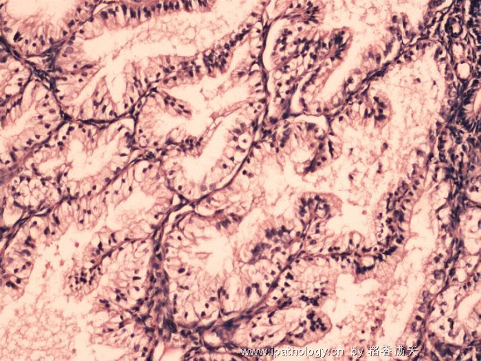
This is not secretory phase endometrium because the stroma is nearly absent and no edema or predecidualization. There are 2 patterns shown in the photos: problematic mucinous lesion (left side of photo 1) and probable background proliferative or weakly proliferative endometrium (right side of photo 1). If what i see here represents the real pictures on slides, i would call this "endometrial intraepithelial neoplasia, EIN) or focal complex hyperplasia. I cannot see nuclear features well, so no comment on cytologic atypia. The last a few photos show back to back glands with negligible stroma, a worrisome sign for adenocarcinoma (would be mucinous adenocarcinoma). Recommend a formal dilatation and curettage (i suppose this is endometrial biopsy?) to exclude malignancy. Be very careful with mucinous epithelium in endometrium. Not sure about endometrial polyp. When this kind of morphology occurs within a endometrial polyp, it is still worrisome for hyperplasia or malignancy. Patient's age is on the young side for endometrial adenocarcinoma, but 37 years old can have EM cancer too. The youngest i have seen is 33 years old, usually very obese or with ovarian problems. Thanks for your analysis! |

朱正龙
This is not secretory phase endometrium because the stroma is nearly absent and no edema or predecidualization. There are 2 patterns shown in the photos: problematic mucinous lesion (left side of photo 1) and probable background proliferative or weakly proliferative endometrium (right side of photo 1). If what i see here represents the real pictures on slides, i would call this "endometrial intraepithelial neoplasia, EIN) or focal complex hyperplasia. I cannot see nuclear features well, so no comment on cytologic atypia. The last a few photos show back to back glands with negligible stroma, a worrisome sign for adenocarcinoma (would be mucinous adenocarcinoma). Recommend a formal dilatation and curettage (i suppose this is endometrial biopsy?) to exclude malignancy.
Be very careful with mucinous epithelium in endometrium.
Not sure about endometrial polyp. When this kind of morphology occurs within a endometrial polyp, it is still worrisome for hyperplasia or malignancy.
Patient's age is on the young side for endometrial adenocarcinoma, but 37 years old can have EM cancer too. The youngest i have seen is 33 years old, usually very obese or with ovarian problems.
