| 图片: | |
|---|---|
| 名称: | |
| 描述: | |
- 颅内顶枕部肿块
-
lfl001200546 离线
- 帖子:2808
- 粉蓝豆:40
- 经验:2808
- 注册时间:2007-02-14
- 加关注 | 发消息
-
lfl001200546 离线
- 帖子:2808
- 粉蓝豆:40
- 经验:2808
- 注册时间:2007-02-14
- 加关注 | 发消息
-
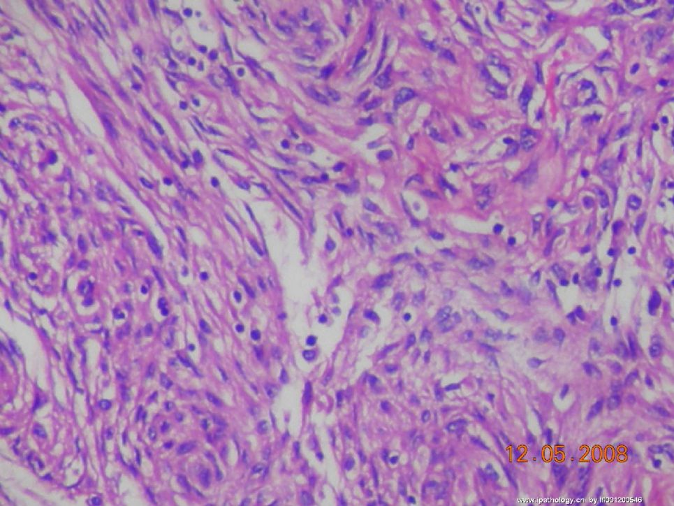
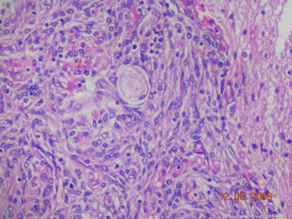
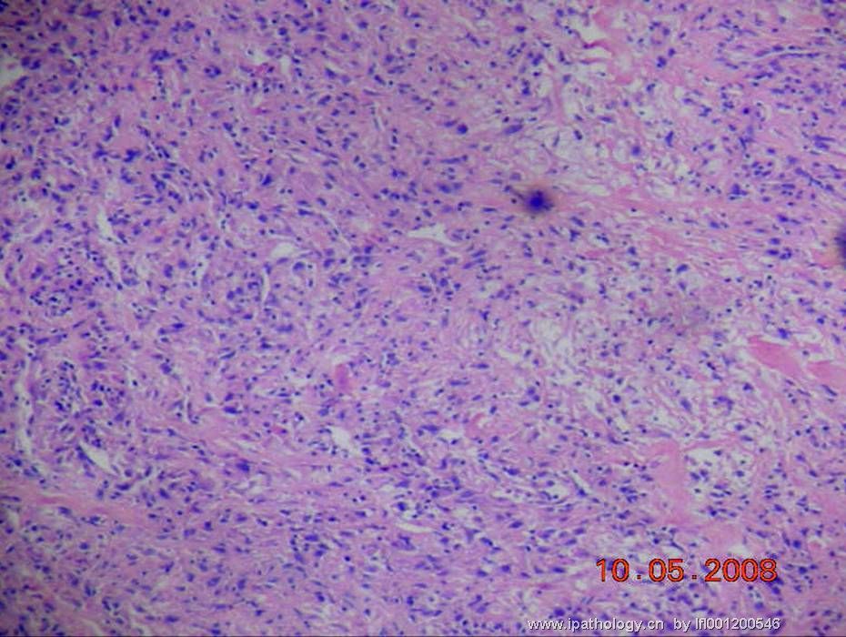
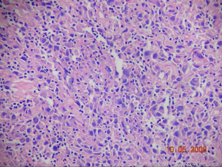
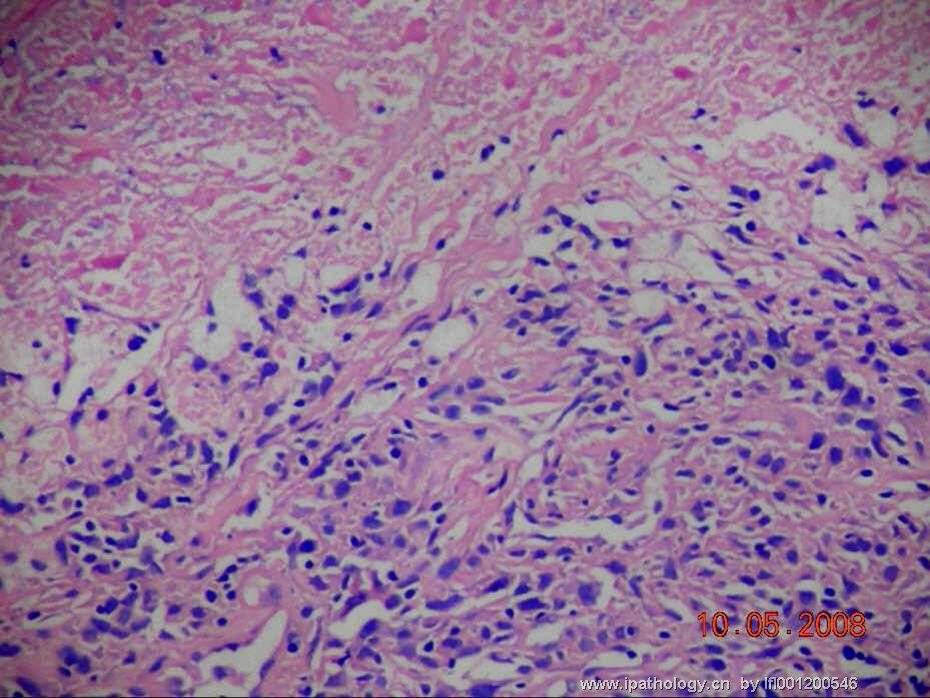
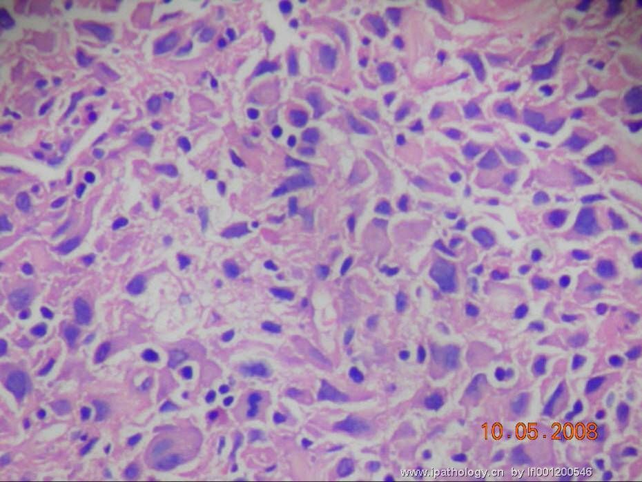
Figures 1 and 2 look like a benign meningioma. 图1-2好象良性脑膜瘤。The history of "destruction of overlying skull bone" is also consistent with meningioma. 颅骨破坏区也是脑膜瘤。Figures 3,4 and 6 contain polygonal cells that do not look like that seen in usual benign meningiomas, but the admixed small lymphocytes are similar to that seen in Figures 1 and 2. 图3、4和图6由多角形性细胞组成不象常见的良性脑膜瘤,但是混有淋巴细胞又类似图1、2。They are not consistent with rhabdoid meningioma, either.这些形态改变也不是横纹肌样脑膜瘤。.I believe they could still be part of the same benign meningioma. 我考虑还是良性脑膜瘤的一部分。Though necrosis is seen in Figure 5, I do not see other atypical features. 虽然图5有坏死,未见有其它非典型性改变特点。The only thing left to do is mitotic count to be sure this is not an atypical meningioma. 应该好好数一下分裂相,以确保其不是非典型性脑膜瘤。
-
Figures 1 and 2 look like a benign meningioma. The history of "destruction of overlying skull bone" is also consistent with meningioma. Figures 3,4 and 6 contain polygonal cells that do not look like that seen in usual benign meningiomas, but the admixed small lymphocytes are similar to that seen in Figures 1 and 2. They are not consistent with rhabdoid meningioma, either..I believe they could still be part of the same benign meningioma. Though necrosis is seen in Figure 5, I do not see other atypical features. The only thing left to do is mitotic count to be sure this is not an atypical meningioma.

聞道有先後,術業有專攻

