共1页/12条首页上一页1下一页尾页
回复:12 阅读:3419
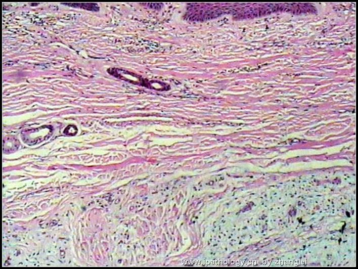
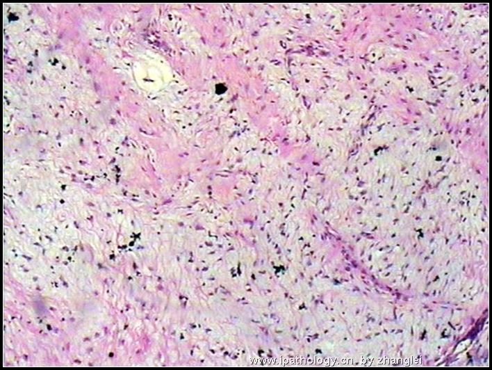
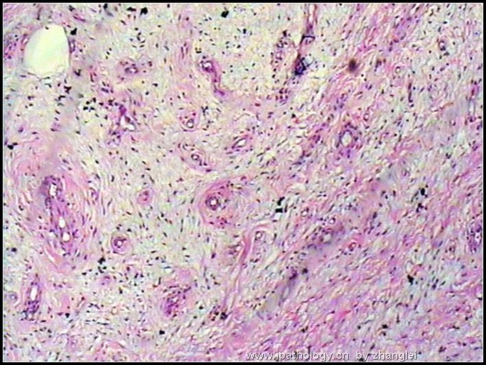
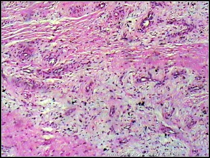
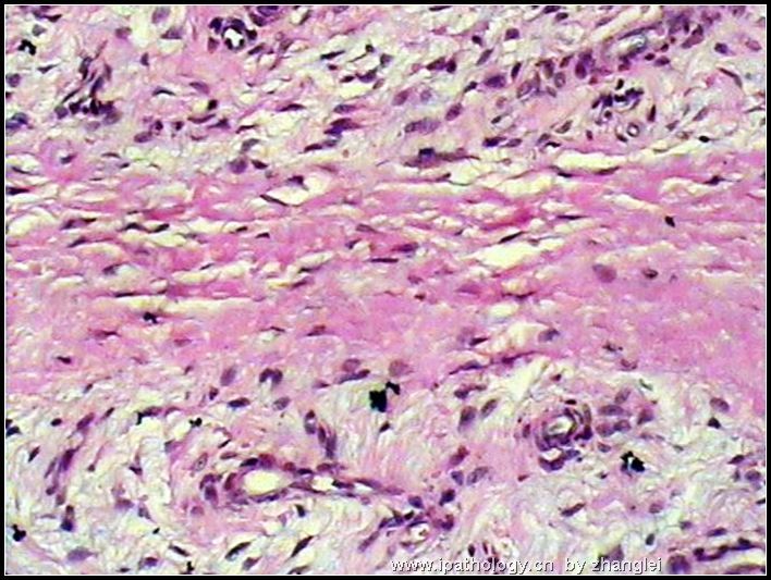
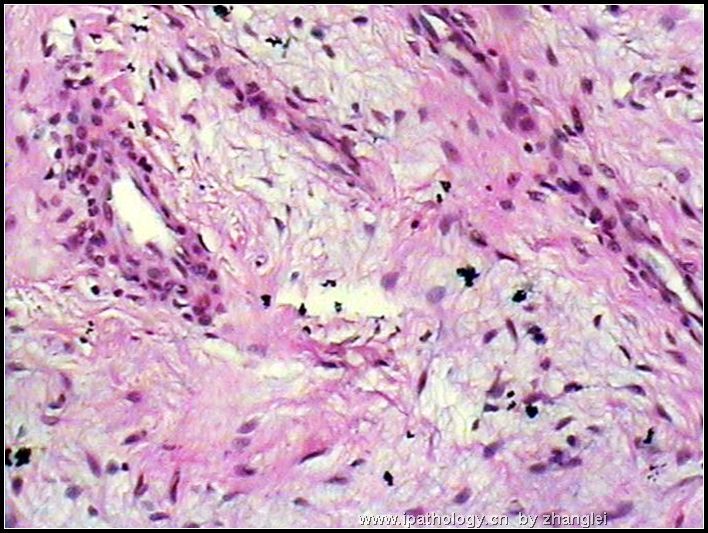
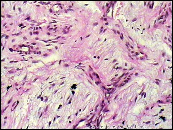
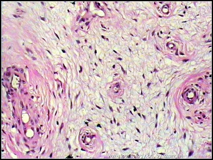
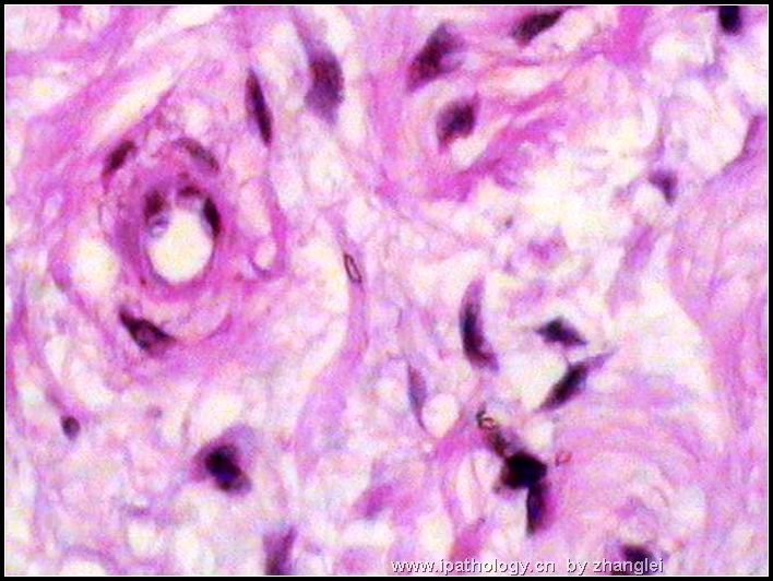
B280右第四趾包块--浅表性血管黏液瘤

zhanglei 离线

帖子:3751
粉蓝豆:2078
经验:3901
注册时间:2006-09-26
加关注  |  发消息
楼主 发表于 2006-11-26 09:18|举报|关注(0)
浏览排序[ 顺序 逆序 楼主 支持 精彩 ]  快捷回复
姓    名: ××× 性别:  男 年龄:  36岁
标本名称:   右第四趾包块
简要病史:  右第四趾无痛性包块10年,逐渐增大。骨组织无破坏。
肉眼检查:  
灰白组织一块7*4*4cm,包裹第四趾骨,切面灰白色,质地中等,有粘液感。
右第四趾包块--浅表性血管黏液瘤图1
名称:图1
描述:图1
右第四趾包块--浅表性血管黏液瘤图2
名称:图2
描述:图2
右第四趾包块--浅表性血管黏液瘤图3
名称:图3
描述:图3
右第四趾包块--浅表性血管黏液瘤图4
名称:图4
描述:图4
右第四趾包块--浅表性血管黏液瘤图5
名称:图5
描述:图5
右第四趾包块--浅表性血管黏液瘤图6
名称:图6
描述:图6
右第四趾包块--浅表性血管黏液瘤图7
名称:图7
描述:图7
右第四趾包块--浅表性血管黏液瘤图8
名称:图8
描述:图8
右第四趾包块--浅表性血管黏液瘤图9
名称:图9
描述:图9
标签:足趾 浅表性血管黏液瘤
本帖最后由 于 2008-01-06 12:11:00 编辑
0
×参考诊断
浅表性血管黏液瘤。

whwwhw 离线

帖子:437
粉蓝豆:7
经验:752
注册时间:2007-12-01
加关注  |  发消息
1 楼    发表于2008-02-13 19:41:00举报|引用
返回顶部 | 快捷回复
以下是引用lllcccyyyy在2008-1-20 11:17:00的发言:

 浅表性血管黏液瘤

0
回复

lllcccyyyy 离线

帖子:53
粉蓝豆:41
经验:53
注册时间:2007-12-12
加关注  |  发消息
2 楼    发表于2008-01-20 11:17:00举报|引用
返回顶部 | 快捷回复
 浅表性血管黏液瘤
0
回复

江边观潮人 离线

帖子:2684
粉蓝豆:524
经验:3338
注册时间:2006-11-12
加关注  |  发消息
3 楼    发表于2007-12-28 23:00:00举报|引用
返回顶部 | 快捷回复
 学习了。谢谢版主!
0
回复
signature

华夏

五月的风 离线

帖子:368
粉蓝豆:19
经验:1359
注册时间:2007-09-19
加关注  |  发消息
4 楼    发表于2007-12-08 23:24:00举报|引用
返回顶部 | 快捷回复
 
0
回复

liguoxia71 离线

帖子:4174
粉蓝豆:3122
经验:4677
注册时间:2007-04-01
加关注  |  发消息
5 楼    发表于2007-12-08 19:58:00举报|引用
返回顶部 | 快捷回复
 表浅血管黏液瘤,学习了。谢谢版主!
0
回复
signature
三人行,必有我师焉,择其善者而从之,其不善者而改之。

wy1992 在线

帖子:4858
粉蓝豆:1
经验:7320
注册时间:2007-06-30
加关注  |  发消息
6 楼    发表于2007-12-07 19:36:00举报|引用
返回顶部 | 快捷回复
 很值得我们学习且时髦的病例!
0
回复
signature

朱正龙

慧眼 离线

帖子:98
粉蓝豆:2
经验:98
注册时间:2006-11-17
加关注  |  发消息
7 楼    发表于2006-12-14 23:39:00举报|引用
返回顶部 | 快捷回复
 
0
回复

zhanglei 离线

帖子:3751
粉蓝豆:2078
经验:3901
注册时间:2006-09-26
加关注  |  发消息
8 楼    发表于2006-12-14 21:01:00举报|引用
返回顶部 | 快捷回复
 范钦和教授会诊:浅表性血管黏液瘤。
0
回复

shihuaiy 离线

帖子:707
粉蓝豆:35
经验:707
注册时间:2006-09-27
加关注  |  发消息
9 楼    发表于2006-11-28 12:12:00举报|引用
返回顶部 | 快捷回复

 发生在指趾,细胞无异型性,我感觉像是表浅肢端纤维粘液瘤(superficial acral fibromyxoma),此瘤2001年首次报道,粘液背景中富于血管。此瘤需要和以下良性肿瘤鉴别:

The differential diagnosis of SAFM encompasses benign or malignant myxoid and
spindle cells tumors showing predilection for distal extremities. Myxoid
neurofibroma is one of the main differential diagnoses as tumor cells in SAFM
often have a neural-like appearance. However, neurofibroma is consistently
positive for S100 protein and does not show the increased vasculature of SAFM.3
Immunostaining for CD34 is not diagnostically helpful as this marker is often
focally positive in neurofibroma. Sclerosing perineurioma, a recently described
tumor of the fingers and palms, is composed of spindled and epithelioid EMA
positive cells arranged in an onion-skin pattern. Although CD34 is expressed in
some perineuriomas, it is usually negative in sclerosing perineurioma.4-6
Superficial angiomyxoma shows a predilection for the head and neck area but can
be encountered in virtually any location. The lesion shows a distinctive
lobulated pattern and is usually poorly demarcated. It is composed of bland
spindle-shaped or stellate cells and neutrophils are characteristically found
within the myxoid matrix. An epithelial component is seen in about one third of
cases.

0
回复
signature
the more we discuss, the more we learn from each other !!

shandongzhang 离线

帖子:804
粉蓝豆:506
经验:1510
注册时间:2006-09-24
加关注  |  发消息
10 楼    发表于2006-11-27 22:52:00举报|引用
返回顶部 | 快捷回复
 看低倍找上皮:除外毛源性黏液瘤(表浅血管黏液瘤)
0
回复

唐伯虎学病理 离线

帖子:364
粉蓝豆:8
经验:364
注册时间:2006-10-02
加关注  |  发消息
11 楼    发表于2006-11-27 22:01:00举报|引用
返回顶部 | 快捷回复
 可能是1,侵袭性血管粘液瘤, 2,韧带样瘤,再传一些低倍和边缘看看,可能明确诊断
0
回复
signature
天天哇哈哈,下班了回家做菜,不要想到取材,那是切菜刀和猪肉,不是标本和取材刀.

amused 离线

帖子:18
粉蓝豆:1
经验:18
注册时间:2006-09-26
加关注  |  发消息
12 楼    发表于2006-11-27 20:35:00举报|引用
返回顶部 | 快捷回复
 一种少见的血管瘤,叫.....?
0
回复
回复:12 阅读:3419
共1页/12条首页上一页1下一页尾页
【免责声明】讨论内容仅作学术交流之用,不作为诊疗依据,由此而引起的法律问题作者及本站不承担任何责任。
快速回复
进入高级回复
您最多可输入10000个汉字,按 "Ctrl" + "Enter" 直接发送
搜索回复/乘电梯 ×
按内容
按会员
乘电梯
合作伙伴
友情链接