共1页/13条首页上一页1下一页尾页
回复:13 阅读:1889
20080306-左颞顶叶肿瘤会诊

shandongzhang 离线

帖子:804
粉蓝豆:506
经验:1510
注册时间:2006-09-24
加关注  |  发消息
楼主 发表于 2008-03-06 20:40|举报|关注(0)
浏览排序[ 顺序 逆序 楼主 支持 精彩 ]  快捷回复
姓    名: ××× 性别:  男 年龄:  72岁
标本名称:  
简要病史: 男72岁,头痛一周,左颞顶肿瘤切除术,临床诊断恶性脑膜瘤,补充强化MRI
诊断?要做哪些工作?谢谢!
肉眼检查:  切面灰红质软,海绵状,部分区域灰白质硬
  • 20080306-左颞顶叶肿瘤会诊图1
    图1
  • 20080306-左颞顶叶肿瘤会诊图2
    图2
  • 20080306-左颞顶叶肿瘤会诊图3
    图3
  • 20080306-左颞顶叶肿瘤会诊图4
    图4
  • 20080306-左颞顶叶肿瘤会诊图5
    图5
  • 20080306-左颞顶叶肿瘤会诊图6
    图6
  • 20080306-左颞顶叶肿瘤会诊图7
    图7
  • 20080306-左颞顶叶肿瘤会诊图8
    图8
  • 20080306-左颞顶叶肿瘤会诊图9
    图9
  • 20080306-左颞顶叶肿瘤会诊图10
    图10
  • 20080306-左颞顶叶肿瘤会诊图11
    图11
  • 20080306-左颞顶叶肿瘤会诊图12
    图12
  • 20080306-左颞顶叶肿瘤会诊图13
    图13
  • 20080306-左颞顶叶肿瘤会诊图14
    图14
  • 20080306-左颞顶叶肿瘤会诊图15
    图15
  • 20080306-左颞顶叶肿瘤会诊图16
    图16
  • 20080306-左颞顶叶肿瘤会诊图17
    图17
  • 20080306-左颞顶叶肿瘤会诊图18
    图18
  • 20080306-左颞顶叶肿瘤会诊图19
    图19
  • 20080306-左颞顶叶肿瘤会诊图20
    图20
标签:
本帖最后由 于 2008-03-07 10:20:00 编辑
0
添加参考诊断
×参考诊断
  

海马 离线

帖子:504
粉蓝豆:12
经验:556
注册时间:2008-08-26
加关注  |  发消息
1 楼    发表于2009-07-22 20:55:00举报|引用
返回顶部 | 快捷回复

谢谢了

0
回复

yourself 离线

帖子:663
粉蓝豆:7132
经验:691
注册时间:2007-06-30
加关注  |  发消息
2 楼    发表于2008-03-06 21:27:00举报|引用
返回顶部 | 快捷回复
 非常好的病例!谢谢zhang老师!组织学上两个特点:一是两种主要细胞,小细胞+胖细胞;一是血管透明变明显,部分区域小细胞围绕血管生长。请zhang老师告知病人年龄,性别,肿瘤具体位置,影像学特点。谢谢!
0
回复

shandongzhang 离线

帖子:804
粉蓝豆:506
经验:1510
注册时间:2006-09-24
加关注  |  发消息
3 楼    发表于2008-03-07 10:20:00举报|引用
返回顶部 | 快捷回复
以下是引用yourself在2008-3-6 21:27:00的发言:

 非常好的病例!谢谢zhang老师!组织学上两个特点:一是两种主要细胞,小细胞+胖细胞;一是血管透明变明显,部分区域小细胞围绕血管生长。请zhang老师告知病人年龄,性别,肿瘤具体位置,影像学特点。谢谢!

已传
0
回复

wfbjwt 离线

帖子:6628
粉蓝豆:163
经验:7418
注册时间:2006-10-08
加关注  |  发消息
4 楼    发表于2008-03-07 19:38:00举报|引用
返回顶部 | 快捷回复
恶性横纹肌样脑膜瘤 
0
回复
signature
嫁人就嫁灰太狼,学习要上华夏网。

yourself 离线

帖子:663
粉蓝豆:7132
经验:691
注册时间:2007-06-30
加关注  |  发消息
5 楼    发表于2008-03-08 07:16:00举报|引用
返回顶部 | 快捷回复
 从影像学及组织学上看,首先考虑横纹肌样脑膜瘤,需免疫组化(EMA,Vim,GFAP,Nestin,NeuN,CK,PR)除外室管膜瘤及胶质或胶质神经元混合性肿瘤。
0
回复

涌泉 离线

帖子:46
粉蓝豆:1
经验:46
注册时间:2008-03-07
加关注  |  发消息
6 楼    发表于2008-03-08 18:07:00举报|引用
返回顶部 | 快捷回复
 恶性横纹肌样脑膜瘤
0
回复

mjma 离线

帖子:703
粉蓝豆:24
经验:789
注册时间:2006-09-28
加关注  |  发消息
7 楼    发表于2008-03-11 11:51:00举报|引用
返回顶部 | 快捷回复
本帖最后由 于 2008-03-11 12:15:00 编辑 This is not an easy case to interpret. Each time I looked at the photos I got some different impression. The neoplasm consists of very loosely arranged cells between large hyalinized blood vessels and amianthoid fibers. The first low power view suggests there is pseudopalisaded tumor necrosis, but high power views do not really confirm necrosis in the loose areas. Though no definite meningothelial whorls or psammoma bodies are seen, there are solid nests of epithelial looking neoplastic cells (especially on photos containing amianthoid fibers) to suggest a meningioma. Was there a history of presurgical embolization of the tumor by an interventional radiologist (which may cause acute infarction with discohesive cells)? MRI images do suggest dural attachment with an enhancing tail, but this does not exclude a surface glioma that adhere to dura. The cellularity is focally very high, suggesting that this is not a benign neoplasm. However, I failed to find mitosis on these uploaded photos. There is prominent perivascular radial arrangement of neoplastic cells, and the loose areas are rimmed by condensed neoplastic cells. It's not an easy case to diagnose without thorough microscopic examination and immunohistochemical stains. My differential diagnoses include meningioma (favored) and glioma. Though the prominent perivascular arrangement and hyalinized blood vessels suggests ependymoma, I would like to see whether the neoplastic cells are uniform or not. Certainly no ependymal canals are seen. If the loose areas are indeed necrosis and mitoses can be found readily with some or much cytologic variation, this would be a WHO grade IV glioblastoma. If cells are fairly uniform without clearcut necrosis, this may be a WHO grade II ependymoma or WHO grade I meningioma. As for the plump epithelioid cells with eosinophilic cytoplasm, I suspect they are just aberrant epithelial differentiation or degenerative change. I do not think this is rhabdoid meningioma. Cells in rhabdoid meningioma are different from the plump eosinophilic cells shown here. Immunohistochemical stains with GFAP, EMA, PR and MIB-1 may help. Interesting case!
0
回复
signature

聞道有先後,術業有專攻

shandongzhang 离线

帖子:804
粉蓝豆:506
经验:1510
注册时间:2006-09-24
加关注  |  发消息
8 楼    发表于2008-03-11 18:04:00举报|引用
返回顶部 | 快捷回复
本帖最后由 于 2008-03-11 18:06:00 编辑

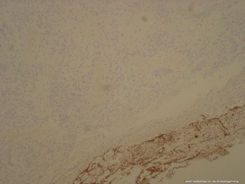
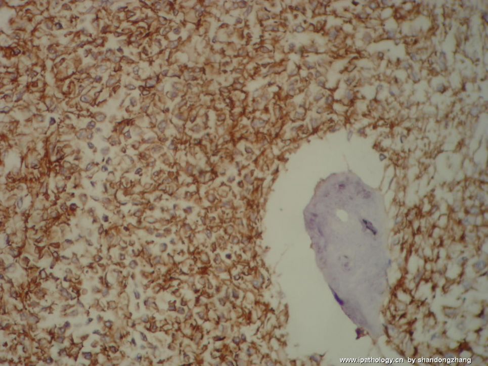
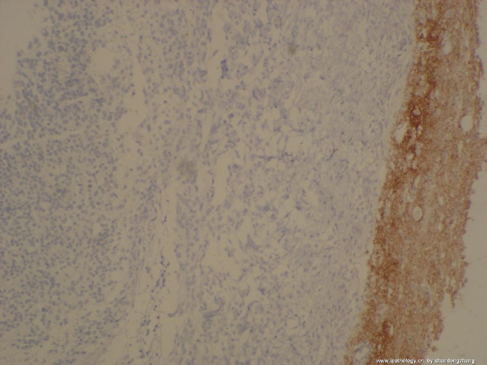
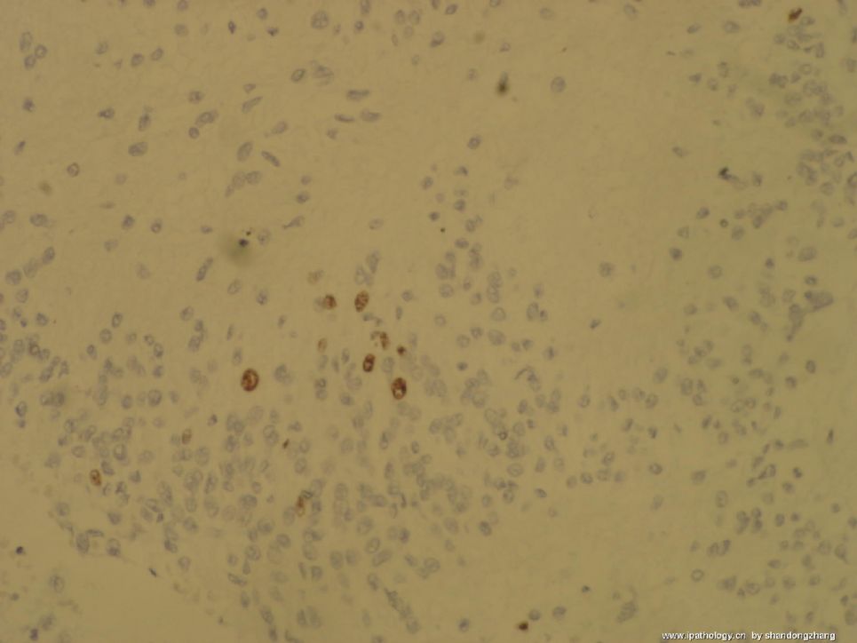
IHC:GFAP\EMA\SYN\KI-67


名称:图1
描述:图1

名称:图2
描述:图2

名称:图3
描述:图3

名称:图4
描述:图4
0
回复

yourself 离线

帖子:663
粉蓝豆:7132
经验:691
注册时间:2007-06-30
加关注  |  发消息
9 楼    发表于2008-03-11 20:57:00举报|引用
返回顶部 | 快捷回复
 meningioma(WHO grade I)
0
回复

mjma 离线

帖子:703
粉蓝豆:24
经验:789
注册时间:2006-09-28
加关注  |  发消息
10 楼    发表于2008-03-12 09:16:00举报|引用
返回顶部 | 快捷回复
This is a WHO grade I meningioma. The areas containing loose neoplastic cells can be very misleading. As much as we rely on immunohistochemical stains nowadays, a good HE section is still very valuable.
0
回复
signature

聞道有先後,術業有專攻

wy1992 在线

帖子:4858
粉蓝豆:1
经验:7320
注册时间:2007-06-30
加关注  |  发消息
11 楼    发表于2008-03-13 12:05:00举报|引用
返回顶部 | 快捷回复
 可以见到明显的横纹肌样细胞,我想请教马老师本例属于何种类型的脑膜瘤
0
回复
signature

朱正龙

shandongzhang 离线

帖子:804
粉蓝豆:506
经验:1510
注册时间:2006-09-24
加关注  |  发消息
12 楼    发表于2008-03-13 15:58:00举报|引用
返回顶部 | 快捷回复
以下是引用wy1992在2008-3-13 12:05:00的发言:

 可以见到明显的横纹肌样细胞,我想请教马老师本例属于何种类型的脑膜瘤

"As for the plump epithelioid cells with eosinophilic cytoplasm, I suspect they are just aberrant epithelial differentiation or degenerative change. I do not think this is rhabdoid meningioma"
0
回复

wy1992 在线

帖子:4858
粉蓝豆:1
经验:7320
注册时间:2007-06-30
加关注  |  发消息
13 楼    发表于2008-03-13 17:24:00举报|引用
返回顶部 | 快捷回复
 谢谢张老师!
0
回复
signature

朱正龙

回复:13 阅读:1889
共1页/13条首页上一页1下一页尾页
【免责声明】讨论内容仅作学术交流之用,不作为诊疗依据,由此而引起的法律问题作者及本站不承担任何责任。
快速回复
进入高级回复
您最多可输入10000个汉字,按 "Ctrl" + "Enter" 直接发送
搜索回复/乘电梯 ×
按内容
按会员
乘电梯
合作伙伴
友情链接