


| 图片: | |
|---|---|
| 名称: | |
| 描述: | |
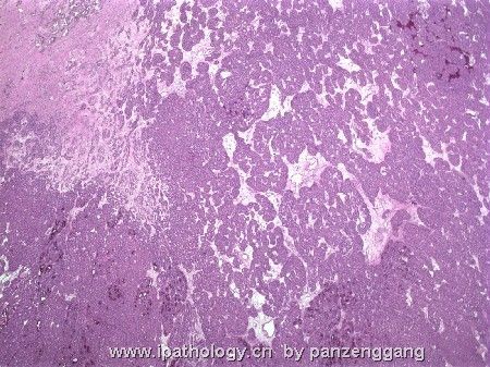
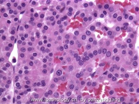
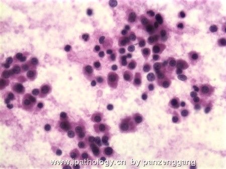
- 肾脏肿瘤—病例1
-
panzenggang 离线
- 帖子:189
- 粉蓝豆:480
- 经验:246
- 注册时间:2008-01-09
- 加关注 | 发消息
-
panzenggang 离线
- 帖子:189
- 粉蓝豆:480
- 经验:246
- 注册时间:2008-01-09
- 加关注 | 发消息
|
1: CK7 2: Colloid iron 3: EMA 4: EM, lower power 5: EM, Higher power | ||||||||||||||||||||||||||||||||||||||||||||||||||||||||||||||||||||||||||||||||
Discussion: Zenggang Pan, MD, PhD. www.enjoypath.com | ||||||||||||||||||||||||||||||||||||||||||||||||||||||||||||||||||||||||||||||||
-
panzenggang 离线
- 帖子:189
- 粉蓝豆:480
- 经验:246
- 注册时间:2008-01-09
- 加关注 | 发消息


