| 图片: | |
|---|---|
| 名称: | |
| 描述: | |
- 左上腹肿块
| 姓 名: | ××× | 性别: | 女 | 年龄: | 44岁 |
| 标本名称: | 左上腹肿块 | ||||
| 简要病史: | 发现左上腹肿块一周。术中见左肾下极巨大肿块25*20* | ||||
| 肉眼检查: | 不整形组织一块18*14* | ||||
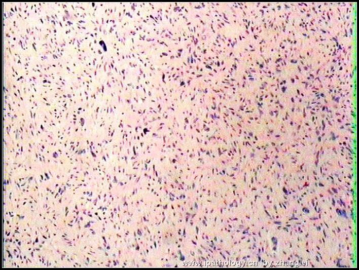
名称:图1
描述:图1
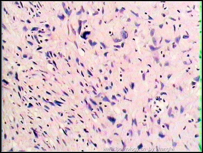
名称:图2
描述:图2
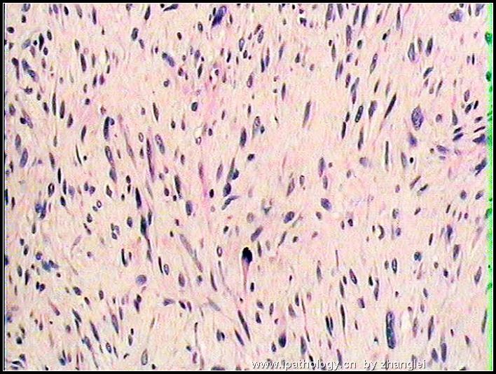
名称:图3
描述:图3
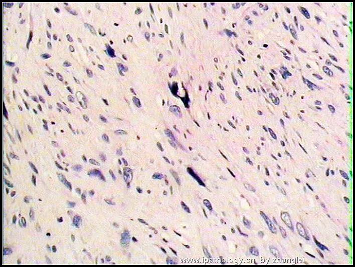
名称:图4
描述:图4
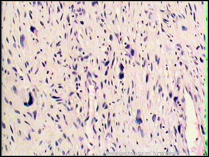
名称:图5
描述:图5
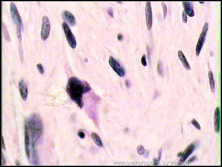
名称:图6
描述:图6
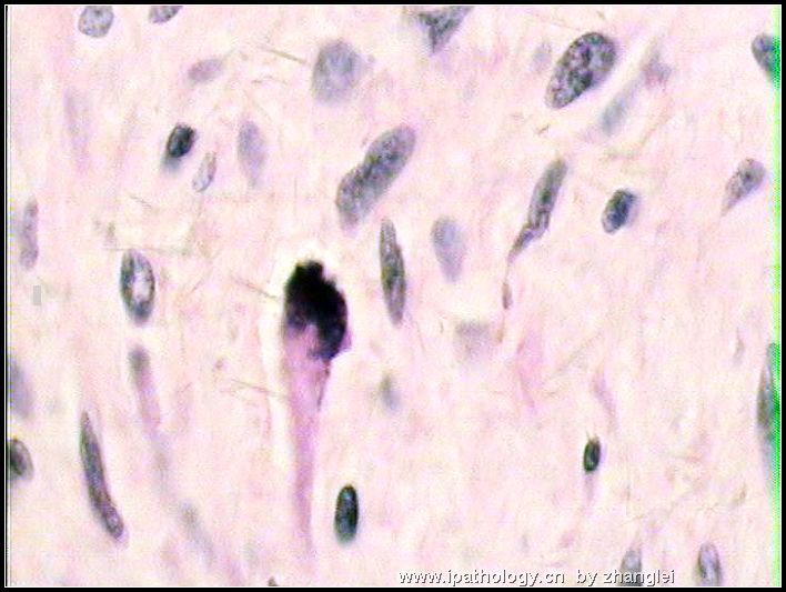
名称:图7
描述:图7
-
本帖最后由 于 2006-11-12 23:17:00 编辑
相关帖子
- • 右侧腹膜后肿块
- • 腹膜后肿物
- • 腹膜后肿物
- • 腹膜后肿瘤
- • 大腿肿物 脂肪肉瘤?
- • 腹膜后肿块
- • 腹膜后肿块(094428)
- • 腹膜后肿物,会诊后公布答案!
- • 腹膜后肿物
- • 腹膜后肿物
-
A retroperitoneal tumor of such large size and histology (spindled cells) has to raise the possibility of dedifferentiated liposarcoma. I urge that any "fat" attached to the tumor be submitted for microscopic examination to detect any feature for a pre-existing and residual low grade liposarcoma. Retroperitoneal liposarcomas are not rare, and are the most common large retroperitoneal tumors in adults. Most of them are low grade or well differentiated liposarcoma (rather than the myxoid or pleomorphic variants of liposarcomas). Microscopically, retroperitoneal well differentiated liposarcomas appear identical to the well circumscribed atypical lipomatous tumors (atypical lipomas) of neck, shoulder, upper back and other regions in extremities. However, retroperitoneal well-differentiated liposarcomas are slow-growing, very large, not encapsulated and very difficult to resect completely due to infiltrative borders.
A minority of retroperitoneal liposarcomas may, either at the time of initial diagnosis or at the time of local recurrence, contain nodule(s) of non-lipogenic sarcomatous elements. Most of these non-lipogenic elements appear like fibrosarcoma or MFH, and often display features of high grade sarcoma. Sometimes, the sarcomatous elements are that of a low grade fibrosarcoma (consisting exclusively of spindled cells). Very rarely, features of high grade osteosarcoma, leiomyosarcoma, chondrosarcoma, rhabdomyosarcoma and malignant peripheral nerve sheath tumor are found. These tumors are known as dedifferentiated liposarcomas. They are presumed to evolve from the pre-existing well differentiated liposarcomas. Unlike well differentiated liposarcomas, dedifferentiated liposarcomas of the retroperitoneum have definite metastatic potential (in 1~18% of cases). No matter what kinds of sarcomatous elements are found in a retroperitoneal dedifferentiated liposarcoma, its clinical behavior is not the same as high grade fibrosarcomas, leiomyosarcomas, osteosarcomas, chondrosarcomas, rhabdomyosarcomas, or MPNST. Dedifferentiated liposarcomas are not unique to the retroperitoneum. Another common location for it is the inguinal area and spermatic cord.

聞道有先後,術業有專攻
| 以下是引用mjma 在2006-11-13 1:56:00的发言: A retroperitoneal tumor of such large size and histology (spindled cells) has to raise the possibility of dedifferentiated liposarcoma. I urge that any "fat" attached to the tumor be submitted for microscopic examination to detect any feature for a pre-existing and residual low grade liposarcoma. Retroperitoneal liposarcomas are not rare, and are the most common large retroperitoneal tumors in adults. Most of them are low grade or well differentiated liposarcoma (rather than the myxoid or pleomorphic variants of liposarcomas). Microscopically, retroperitoneal well differentiated liposarcomas appear identical to the well circumscribed atypical lipomatous tumors (atypical lipomas) of neck, shoulder, upper back and other regions in extremities. However, retroperitoneal well-differentiated liposarcomas are slow-growing, very large, not encapsulated and very difficult to resect completely due to infiltrative borders. |
非常同意 马教授的观点,此例从细胞排列、形态以及间质特点来看,都需要首先考虑去分化脂肪肉瘤的可能。免疫组化在区分去分化脂肪肉瘤与外周神经肿瘤方面好像并没有太大帮助,多取材找相对典型区域可能帮助更大。最近有文献报道MDM-2以及CDK-4在去分化脂肪肉瘤阳性率较高,我们用的结果也还可以。就此例形态来讲主要是去分化脂肪肉瘤、MFH、MPNST之间的鉴别,但有文献报道腹膜后MFH多数可能都是去分化脂肪肉瘤,如果此结论可信的话,这例主要是去分化脂肪肉瘤和MPNST之间的鉴别了。
建议再多取材找找有无相对典型一些的区域。再追问一下以往有无手术史,如果有的话可以复查一下原切片。

- the more we discuss, the more we learn from each other !!


