| 图片: | |
|---|---|
| 名称: | |
| 描述: | |
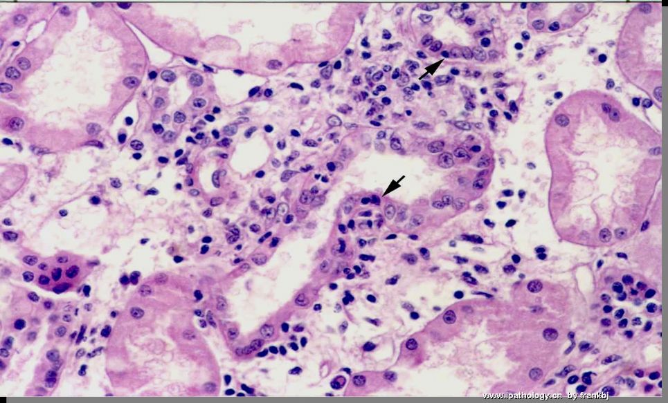
- 肾脏移植半年后穿刺
-
本帖最后由 于 2009-07-13 21:52:00 编辑
The tubule in the center of the photo shows intraepithelial mononuclear cell infiltration. That is tubulitis. Because it is longitudinal section of that tubule, the tubulitis should be graded based on X mononuclear cells/10 tubular cells. There are more than 20 tubular epithelial cells in that tubule with 11 or 12 infiltrating mononuclear cells. The final grading would be t2 (5-6/10 tubular cells, moderate tubulitis). The other tubule close to the right upper corner demonstrates 4 infiltrating mononuclear cells. It should be graded as t1. If the interstitium is more than 25% inflamed (i2), these findings will be enough to be diagnosed of acute T-cell-mediated rejection, Banff type 1A (i2, t2).
-
zhongshihua 离线
- 帖子:1608
- 粉蓝豆:0
- 经验:1651
- 注册时间:2006-09-11
- 加关注 | 发消息


