| 图片: | |
|---|---|
| 名称: | |
| 描述: | |
- 太罕见了
| 姓 名: | ××× | 性别: | 年龄: | ||
| 标本名称: | |||||
| 简要病史: | |||||
| 肉眼检查: | |||||
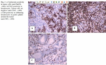
| A rare gastric neoplasm: gastric medullary carcinoma A 57-year-old woman was admitted to the hospital with fatigue, sweating, dyspepsia and abdominal pain radiating to her back for a few months. On examination, she had epigastric tenderness without defense or rebound. She had no comorbid disease. Upper gastrointestinal biopsy revealed undifferentiated adenocarcinoma, and there was only antral wall thickening on computerized tomography. She had a subtotal distal gastrectomy. An ulcerated tumor having expanded margins limited in the submucosa with dense lymphocytic infiltrate was detected microscopically (Fig. 1a). Sheets of polygonal cells with vesicular nuclei, abundant cytoplasm and marked infiltrating lymphocytic response were observed (Fig. 1b). Tumor cells showed an organoid and trabecular architecture. Immunohistochemically tumor cells were stained positively with cytokeratin (Fig. 2a), whereas dense lymphocytic infiltration around the tumor cells was stained positively with CD45 (Fig. 2b). CD3 was positive in infiltrating lymphocytes (Fig. 2c). In situ hybridization analysis of Epstein–Barr virus (EBV) latencyassociated RNA (EBER) was negative. No metastasis was found in regional lymph nodes. The case was diagnosed as gastric medullary carcinoma with no EBV association. We did not consider any adjuvant treatment, since the disease stage was T1N0M0, according to TNM staging system. She has had no relapse until now with 10 months of follow-up. |
标签:
×参考诊断


