| 图片: | |
|---|---|
| 名称: | |
| 描述: | |
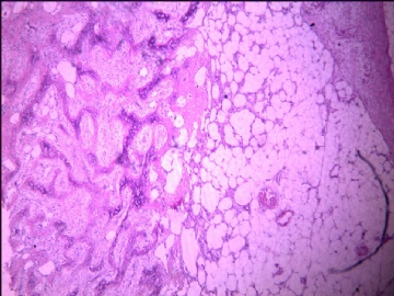
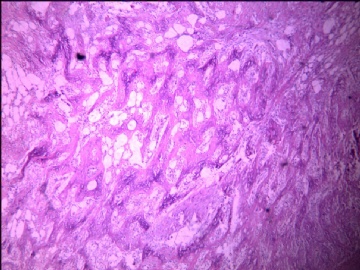
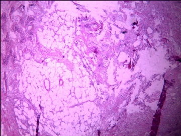
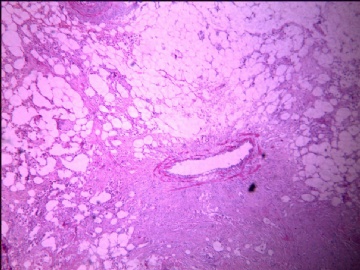
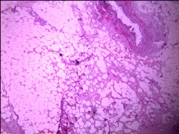
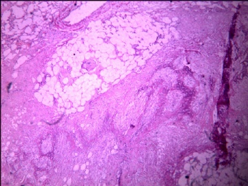
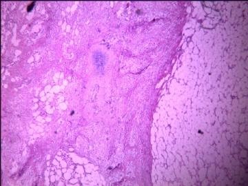
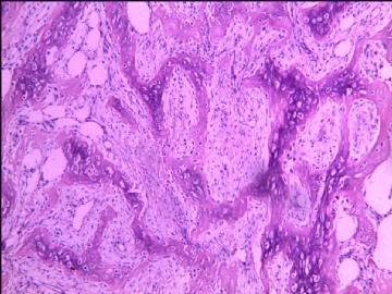
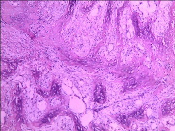
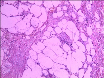
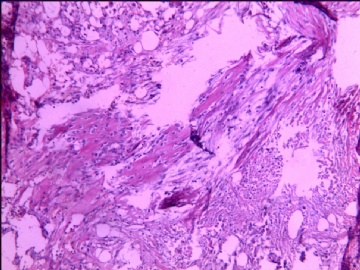
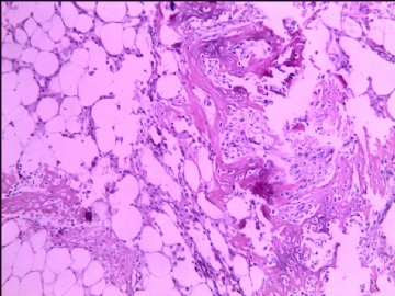
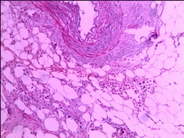
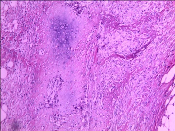
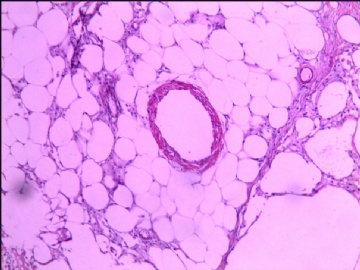
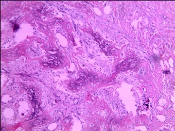
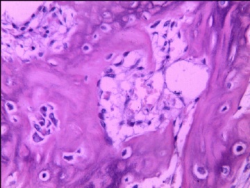
- 回肠肠系膜肿物(少见病例)
| 姓 名: | ××× | 性别: | 男 | 年龄: | 74 |
| 标本名称: | 回肠肠系膜肿物 | ||||
| 简要病史: | 阑尾术后17天肠梗阻行肠管切除术,肠系膜扪及4*1厘米不整形硬结。 | ||||
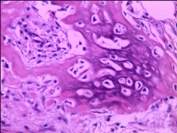
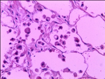
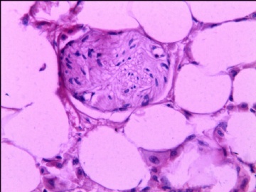
| 肉眼检查: | 脂肪组织中见多处不规则形灰白质硬区,与周围脂肪无明显分界,无包膜。各个病变区之间有较长间隔。 | ||||
您的诊断:
1、良性间叶瘤
2、恶性间叶瘤
3、腹腔内骨化性肌炎
4、骨化性脂膜炎
5、pecoma伴骨化
6、肠壁浆膜面骨化
7、其他
-
本帖最后由 于 2011-03-22 09:45:00 编辑
-
本帖最后由 于 2011-03-05 21:08:00 编辑
非常罕见的腹腔假肉瘤样病变:文献中仅28例报道.
上海长海医院最近有一例报道[1].
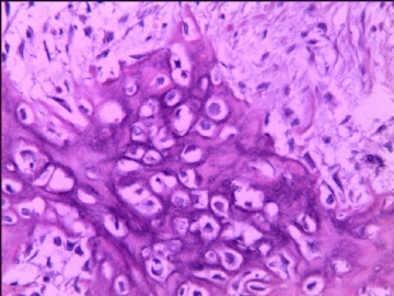
诊断:肠系膜异位骨化 Heterotopic mesenteric ossification[8],也有学者用 "腹腔骨化性肌炎", 但是本人认为称为腹腔骨化性肌炎不妥.
"肠系膜异位骨化"的名称 1999年由Wilson 和 Rosai[15]等命名.
此外腹膜也可有软骨分化[13].请参考楼下文献:
由衷感谢学浅老师与我们分享罕见病例!

- xljin8
-
本帖最后由 于 2011-03-05 21:07:00 编辑
| 以下是引用xljin8在2011-3-5 20:42:00的发言: 1.Surg Today. 2011 Jan;41(1):137-40. Epub 2010 Dec 30. Early postoperative heterotopic omental ossification: report of a case. Shi X, Zhang W, Nabieu PF, Zhao W, Fu C. Department of Colorectal Surgery, Changhai Hospital, Second Military Medical University, No 168 Changhai Road, Shanghai 200433, PR China.Heterotopic mesenteric ossification (HMO) is an uncommon disorder that may sometimes be misdiagnosed. It can cause bowel or intestinal perforation, which may also lead to serious complications or even death. Heterotopic bone formation in the omentum, which is called heterotopic omental ossification (HOO) and is one type of HMO, is considered to be an exceedingly rare event. To our knowledge,about 29 casesof HMO have been reported in previous studies, of which three were HOO. We herein describea case of HOO occurring in a 39-year-old Chinese man with no medical history of abdominal surgery. He underwent a left hemicolectomy, which was performed for the treatment of descending colon adenocarcinoma. Two weeks later, he developed a small bowel obstruction associated with multiple foci of heterotopic bone formation within the omentum. He therefore underwent a second surgical procedure for adhesiolysis and a partial omentectomy. The postoperative course was uneventful. He is still alive and disease-free 16 months later.2.Obes Surg. 2010 Sep;20(9):1312-5. Epub 2010 Feb 2.Heterotopic mesenteric ossification following gastric bypass surgery: case series and review of literature. Yushuva A, Nagda P, Suzuki K, Llaguna OH, Avgerinos D, Goodman E. Department of Surgery, Beth Israel Medical Center, Albert Einstein College of Medicine, First Avenue at 16th Street, New York, NY 10003, USA. ayushuva@yahoo.com Heterotopic mesenteric ossification (HMO) is a rare entity with few cases reported in the world literature. We report two cases. Both patients underwent an open gastric bypass with Roux-en-Y reconstruction procedure for morbid obesity and subsequently presented with gastrointestinal fistulae associated with HMO. 3.J Trauma. 2008 Dec;65(6):1567.Extensive heterotopic mesenteric ossification after penetrating abdominal trauma. Como JJ, Yowler CJ, Malangoni MA. Department of Surgery, MetroHealth Medical Center, Case School of Medicine, Cleveland, Ohio 44109, USA. jcomo@metrohealth.org 4.Clin Nucl Med. 2008 Jul;33(7):496-9. Imaging characteristics of heterotopic mesenteric ossification on FDG PET andTc-99m bone SPECT. Deryk S, Goethals L, Vanhove C, Geers C, Vandenbroucke F, Hove KV, Bossuyt A, Lahoutte T. Department of Nuclear Medicine, UZ Brussels, Brussels, Belgium. sylviaderyk@hotmail.com A CT scan of a 69-year-old male patient, performed for staging of suspected lung carcinoma, incidentally showed an irregular lesion of 10 cm in the upper abdomen. Further investigation using FDG-PET showed only moderately increased glucose metabolism, whereas Tc-99m MDP SPECT revealed intense osteoblastic activity inside the lesion. A CT-guided biopsy was performed and histologic analysis established the diagnosis of heterotopic mesenteric ossification. This pathology is rare and mostly diagnosed when it is complicated by small bowel obstruction.5.J Formos Med Assoc. 2007 Feb;106(2 Suppl):S32-6. Heterotopic mesenteric ossification after total colectomy for bleedingdiverticulosis of the colon--a rare case report. Lai HJ, Jao SW, Lee TY, Ou JJ, Kang JC. Division of Colorectal Surgery, Department of Surgery, Tri-Service General Hospital, Taipei, Taiwan. 6.Heterotopic bone formation within an abdominal incision is a rare sequela of abdominal surgery. Only a few previous reports have noted heterotopic ossification in the mesentery of the small intestine and peri-ileostomy. Here, we report the case of a 60-year-old man who underwent emergent laparotomy and total colectomy with end ileostomy and developed this condition 1 month postoperatively. Heterotopic ossification in the peri-ileostomy tissue caused stenosis of the ileostoma. Laparotomy for re-anastomosis due to a large bone formation at an abdominal midline scar is very difficult and results in a massive abdominal wall defect. Therefore, we used a lower transverse incision to avoid the site of bone formation and resected the terminal ileum with its ossified mesentery. Then, we successfully carried out an anastomosis between the ileum and the rectum. The possible pathogenesis is a metaplastic mechanism ofdifferentiation of immature multipotent mesenchymal cells. Our case provides the experience of treatment and new perspective on currently held hypotheses of heterotopic bone formation.7.Int J Surg Pathol. 2006 Jan;14(1):37-41. Intraabdominal myositis ossificans: a report of 9 new cases. Zamolyi RQ, Souza P, Nascimento AG, Unni KK. Division of Anatomic Pathology, Mayo Clinic, Rochester, MN 55905, USA.Intraabdominal myositis ossificans (IMO) is a rare benign disorder characterized by reactive bone formation in intraabdominal soft tissue that should be distinguished from a malignant condition. We retrospectively searched our patient records and report 9 new cases of IMO. The lesions occurred in 7 men and 2 women with a mean age of 50 years (range, 24--76 years), 5 of whom had previous abdominal surgery. Histologically, all the cases were similar, consisting of a reactive mesenchymal process in adipose tissue. Mitosis was observed, but with no atypical forms, and the lesions lacked malignant cytologic features. IMO is an uncommon benign lesion that develops relatively rapidly. The pathogenesis is related to intraabdominal surgical procedures, but the exact mechanism remains to be determined.8. Patel RM, Weiss SW, Folpe AL.
experience with 6 additional cases, all of which were referred to us with a diagnostic consideration of extraskeletal osteosarcoma (EO) or "sarcoma" and emphasize features which distinguish HMO from EO. Six intraabdominal lesions coded as "heterotopic mesenteric ossification," "ossifying pseudotumor," or"reactive myofibroblastic proliferation with ossification" were retrieved from our consultation files. Clinical follow-up information was obtained. Lesions occurred exclusively in males, with a mean patient age of 49 years (range, 22-72 years). The tumors occurred in the mesentery (N = 4), omentum (N = 1), or both (N= 1) and were preceded by significant abdominal surgery (4 cases) or trauma (1case) in all but 1 case. Five patients presented with bowel obstruction and 1 with abdominal sepsis. Tumors were difficult to precisely measure; the mean size of the resection specimens was 11.8 cm (range, 3.5-20 cm). Grossly, the tumors resembled fat necrosis and often cut with a gritty sensation. Microscopically,all lesions demonstrated an exuberant, reactive (myo)fibroblastic proliferation resembling nodular fasciitis, with extensive hemorrhage and fat necrosis. All tumors produced abundant bone and osteoid, often "lace-like," and 2 contained cartilage. The proliferating (myo)fibroblasts, osteoblasts, and chondroblasts were mitotically active but cytologically bland. Follow-up (4 cases; mean, 47.3 months; range, 5-120 months) showed 3 patients alive without disease and 1 dead of unrelated causes. One case was recent. HMO is a distinct intraabdominal
ossifying pseudotumor that typically occurs in males, almost always after surgery or abdominal trauma, and frequently presents with symptoms of intestinal obstruction. This clinical history, presence of clearly reactive zones resembling nodular fasciitis, thick osteoid, and absence of nuclear atypia, necrosis, and atypical mitotic figures allow the distinction of HMO from its most important morphologic mimic, EO.
Tonino BA, van der Meulen HG, Kuijpers KC, Mallens WM, van Gils AP.Department of Radiology, Ziekenhuis Leyenburg, Den Haag, The Netherlands. btonino@yahoo.comHeterotropic mesenteric ossification is a rare entity. Only a few cases have been described in the literature. We report a case of heterotropic mesenteric ossification in a patient who underwent several laparotomies, after suffering from multiple gunshot wounds. We discuss the radiographic findings of this disease that can easily be misdiagnosed, and review the literature.10. Int J Surg Pathol. 2004 Oct;12(4):407-9. Heterotopic mesenteric ossification ("intraabdominal myositis ossificans''): a case report.Bovo G, Romano F, Perego E, Franciosi C, Buffa R, Uggeri F.Division of Pathology, San Gerardo Hospital, II University of Milan-Bicocca, Italy. Heterotopic ossification has been reported only rarely within the abdominal cavity, specifically in a mesenteric location (heterotopic mesenteric ossification). We describe the case of a 76-year-old man with no history of previous surgery who developed small bowel obstruction associated with multiple foci of heterotopic bone formation within the small bowel mesentery. He underwent small bowel and mesentery resection and is disease-free 9 months later. 11. Rontgenpraxis. 2003;55(3):99-102. [Mesenteric ossification in CT indicates sclerosing peritonitis in chronicbacterial infection and pancreatitis]. [Article in German] Kirchner J, Kickuth R, Stein A, Kirchner EM.Abteilung für Diagnostische und Interventionelle Radiologie, Klinikum Niederberg Velbert. Sclerosing peritonitis already has been described as a serious complication ofthe continuous ambulatory peritoneal dialysis. But different other affections of the peritoneum such as chronic bacterial peritonitis and pancreatitis may result in sclerosing peritonitis, too. The symptom is characterised by thickened small bowel walls and peritoneal membranes as well as peritoneal calcifications which can be shown in computed tomography. We demonstrate two cases of peritoneal ossifications due to peritonitis and pancreatitis. 12. Gastroenterol Clin Biol. 2004 Feb;28(2):188-9. [Heterotopic mesenteric ossification: a rare cause of postoperative occlusion]. [Article in French] Compérat E, De Saint-Maur PP, Kharsa G, Fléjou JF.Service d'Anatomie et Cytologie Pathologiques, Hôpital Saint-Antoine, AP-HP, 184, rue du Faubourg Saint-Antoine, 75571 Paris Cedex 12. Intraabdominal heterotopic ossification is a very rare lesion, especially in the mesentery. In all reported series, there was a history of traumatic injury of the abdomen. We report two cases of heterotopic mesenteric ossification occurring after laparotomy in adults. 13. Mod Pathol. 2002 Jul;15(7):777-80. Cartilaginous differentiation in peritoneal tissues: a report of two cases and a review of the literature.Fadare O, Bifulco C, Carter D, Parkash V.Department of Pathology, Yale University School of Medicine, New Haven,Connecticut 06520, USA.Two cases of cartilaginous differentiation of the peritoneum not associated with an intraabdominal malignancy are described. This is the first detailed report of cartilaginous metaplasia of the peritoneum. The patients were female, ages 53 (Patient 1) and 77 years (Patient 2). Prior medical histories were significant for a culdotomy (to drain pelvic abscesses associated with pelvic inflammatory disease) in Patient 1 and for an open abdominal surgery in Patient 2. The peritoneal lesions were incidental findings in both cases. In Patient 1, surgery was performed for a septated ovarian cyst; the other patient underwent surgery to relieve obstructive bowel symptoms. In Patient 1, multiple firm, white lesions ranging from 2.0 to 7.0 mm were present on the serosal surfaces and the mesenteries of the small and large bowel. In Patient 2, a single firm, white lesion measuring 2 cm in maximum dimension was removed from the mesentery of the ileum. Microscopically, the lesions consisted of small nodules of mature hyaline cartilage surrounded by nondescript fibrous tissue and covered by mesothelium. There was no foreign body giant cell reaction, inflammation, or other reactive changes in the surrounding adipose tissue. These may represent metaplastic lesions of the secondary mullerian system, or a unique peritoneal response to previous surgical manipulation. Alternatively, these may represent benign neoplastic lesions (chondroma) of the submesothelium.14. Pathologica. 2000 Oct;92(5):331-4. [Heterotopic mesenteric ossification. Description of a case].[Article in Italian] Marucci G, Spitale LS, Piccinni DJ. Dipartimento di Oncologia, Sezione di Anatomia, Istologia e Citologia Patologica M. Malpighi, Università di Bologna, Ospedale Bellaria, Argentina.We describe a case of heterotopic mesenteric ossification presented in a 25 year-old male who underwent laparotomy for a fire-gun injury. Two weeks later he experienced small bowel obstruction and for this reason he has been operated five times with removal of segments of small bowel. Now, nine months later, he needs ileostomy to avoid another obstruction.Heterotopic mesenteric ossification ('intraabdominal myositis ossificans'): report of five cases.
Wilson JD, Montague CJ, Salcuni P, Bordi C, Rosai J.Department of Pathology, Memorial Sloan-Kettering Cancer Center, New York, New York 10021, USA. Intraabdominal heterotopic ossification is a very uncommon disorder. We report five new cases, review the previous literature, and discuss the clinical and pathologic features of these lesions. The clinical features of the current cases and of those previously reported are remarkably similar. All patients were middle-aged to elderly men (range, 43-80 years; mean, 61 years) who had small bowel obstruction associated with heterotopic bone formation in the small bowel mesentery, often after one or more abdominal operations. In one case, an initial diagnosis of extraosseous osteosarcoma was considered. This unusual reactive process shares many of the clinical and pathologic features of myositis ossificans, as classically described in somatic soft tissues. We propose to designate this condition heterotopic mesenteric ossification.16. Am J Gastroenterol. 1992 Feb;87(2):230-3. Mesenteritis ossificans. Heterotopic ossification within the small-bowel mesentery.Myers MA, Minton JP.Department of Surgery, Ohio State University College of Medicine, Columbus 43210. Heterotopic bone formation has been previously noted in abdominal laparotomy scars, but the presence of ectopic bone within the peritoneum is extremely rare. Our patient had recurrent formation of heterotopic bone involving the abdominal wall, peritoneum, and small-bowel mesentery. The features of various types of ectopic calcification are discussed, and several theories concerning the pathogenesis and treatment of heterotopic ossification are examined. |

- xljin8
-
zhenshijian 离线
- 帖子:1069
- 粉蓝豆:91
- 经验:1364
- 注册时间:2008-04-20
- 加关注 | 发消息

