
| 图片: | |
|---|---|
| 名称: | |
| 描述: | |
- Pancreatic lesion
-
panzenggang 离线
- 帖子:189
- 粉蓝豆:480
- 经验:246
- 注册时间:2008-01-09
- 加关注 | 发消息
| 姓 名: | ××× | 性别: | Male | 年龄: | 66 |
| 标本名称: | Pancreatic lesion | ||||
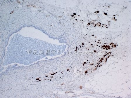
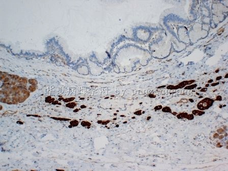
| 简要病史: | A 66 years male had an intraductal papillary mucinous neoplasm of pancreas (IPMN). Scattered atypical cells were noted in or surrounding the IPMN. | ||||
| 肉眼检查: | |||||

名称:图1
描述:图1

名称:图2
描述:图2

名称:图3
描述:图3

名称:图4
描述:图4

名称:图5
描述:图5

名称:图6
描述:图6

名称:图7
描述:图7

名称:图8
描述:图8

名称:图9
描述:图9

名称:图10
描述:图10
标签:
×参考诊断
Benign entrapped islet cells
-
panzenggang 离线
- 帖子:189
- 粉蓝豆:480
- 经验:246
- 注册时间:2008-01-09
- 加关注 | 发消息
-
本帖最后由 于 2011-03-14 08:05:00 编辑
Final diagnosis: Benign entrapped neuroendocrine cells (islet cells) due to chronic pancreatitis associated with IPMN.
These islet cells show atypia with hyperchromasia and arrangement in small nests, single lines or individual cells, which may mimic invasive carcinoma. Similar changes can be noted in adjacent reactive lobules.
The following picture shows a transition from residual islets (right side), to small nests (middle), and to individual cells (left).


