| 图片: | |
|---|---|
| 名称: | |
| 描述: | |
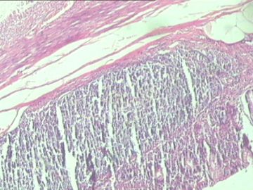
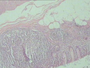
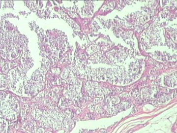
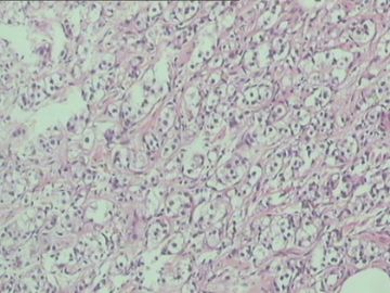
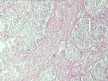
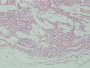
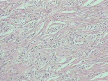
- 请各位老师指点:阑尾
-
本帖最后由 于 2011-02-01 20:09:00 编辑
有关文献:
1. Rossi G, Nannini N, Bertolini F, Mengoli MC, Fano R, Cavazza A.
Clear cell carcinoid of the appendix: an uncommon variant of lipid-rich neuroendocrine tumor with a broad differential diagnosis.
Endocr Pathol. 2010 Dec;21(4):258-62.
Policlinico of Modena, Modena, Italy. rossi.giulio@policlinico.mo.it
The designation of clear cell/lipid-rich refers to an unusual variant of
neuroendocrine tumor ("carcinoid") described in several organs, but only recently observed in the appendix. In this study, we report the morphologic, immunohistochemical, and ultrastructural features of an incidentally discovered appendiceal clear cell/lipid-rich carcinoid in a 32-year-old man without any evidence of von Hippel-Lindau disease. Differential diagnosis with mimicking neoplastic and non-tumor lesions, epidemiology, and clinical behavior of this exceedingly rare variant of carcinoid of the appendix are also discussed.
2. La Rosa S, Finzi G, Puppa G, Capella C.
Lipid-rich variant of appendiceal well-differentiated endocrine tumor(carcinoid).
Am J Clin Pathol. 2010 May;133(5):809-14.
Servizio di Anatomia Patologica, Ospedale di Circolo, Viale Borri 57, 21100
Varese, Italy.
Well-differentiated endocrine tumors (WDETs) of the appendix show characteristic morphologic features, including proliferation of cells with finely granulated eosinophilic cytoplasm. However, clear cell WDETs, which can present a diagnostic challenge, have been occasionally described, but it is unknown whether they represent a morphologic variant with distinct clinicopathologic features. Moreover, the clear cell appearance of the cytoplasm has never been explained. We studied 13 appendiceal WDETs composed of clear cells, which showed an immunophenotype identical to that of conventional appendiceal WDETs. Ultrastructural examination demonstrated abundant lipid accumulation. Patient survival was excellent and equal to that of conventional appendiceal WDETs. These neoplasms, which represent a lipid-rich variant of appendiceal WDETs, do not have different relevant clinical implications compared with conventional WDETs, but it is important to know of their existence for the differential diagnosis with more aggressive neoplasms, including goblet cell carcinoids and appendiceal metastases from clear cell carcinomas.
3. Chetty R, Serra S.
Lipid-rich and clear cell neuroendocrine tumors ("carcinoids")of the appendix: potential confusion with goblet cell carcinoid.
Am J Surg Pathol. 2010 Mar;34(3):401-4.
Department of Pathology, Laboratory Medicine Programme, University Health Network/University of Toronto, Toronto, Canada. runjan.chetty@uhn.on.ca
The so-called clear cell change has been described in neuroendocrine tumors at several locations. Those associated with von Hippel Lindau disease are pathognomonically "clear" and the cytoplasmic appearance has been ascribed to intracytoplasmic lipid. However, lipid has not been demonstrated in all cases of clear cell carcinoid tumors. Such variants have not been described in carcinoid tumors of the appendix and cases with a prominent proportion of clear or more correctly, lipid-rich cytoplasm may bear a superficial resemblance to goblet cell carcinoid and/or signet ring adenocarcinoma. Seven cases, in 5 females and 2 males ranging in age from 22 to 65 years, were noted to have a population of lipid-rich and vacuolated clear cells accounting for 25% or more of the tumor population. The carcinoid tumors were incidental in all cases with 4 of patients presenting with appendicitis, 2 with concomitant mucinous cystadenocarcinomas of the appendix and 1 with an adenocarcinoma of the ascending colon. Morphologically, the tumors had a nested and trabecular pattern and were composed of an admixture of microvesicular and clear lipid-rich cells. There were no mitoses, areas of necrosis of lymphovascular invasion and all cases extended to the mesoappendix. All cases were positive for synaptophysin, chromogranin, and serotonin but negative for inhibin. Three cases were examined ultrastructurally, and showed the presence of intracytoplasmic lipid and neurosecretory granules. None of the patients have shown evidence of recurrent disease. The importance of recognizing this variant of carcinoid tumor in the appendix is to avoid confusion with goblet cell carcinoid tumors with or without a signet ring adenocarcinoma. The presence of multi-vacuolated, foamy and clear cells, some resembling signet ring or goblet cells, in otherwise classic carcinoid tumors is rare but should be considered in this context in the appendix.

- xljin8
