


| 图片: | |
|---|---|
| 名称: | |
| 描述: | |
- “浸润性微乳头状癌”诊断标准和生物学意义(附乳腺 尿路 结直肠 胆管 壶腹部 胰 肺和卵巢 的相关文献摘要和..
| 姓 名: | ××× | 性别: | 年龄: | ||
| 标本名称: | |||||
| 简要病史: | |||||
| 肉眼检查: | |||||
近年来病理医生对癌组织中出现的“微乳头状结构 (micropapillary pattern)”倍加关注,例如肺腺癌、尿路上皮癌、结肠癌、乳腺癌等都有较多文献报道。大多研究认为“微乳头状癌”与预后有关。因此,如何在形态学上把握诊断“微乳头状癌”非常重要,漏了可能导致治疗不足;相反,诊断过了可能导致过度治疗。请感兴趣的网友发表意见。
以下是"乳腺浸润性微乳头状癌"的有关病例,请参阅及发表意见, 谢谢!
http://www.ipathology.cn/forum/forum_display.asp?keyno=342819
http://www.ipathology.cn/forum/forum_display.asp?keyno=278637
http://www.ipathology.cn/forum/forum_display.asp?keyno=342108
http://ipathology.cn/forum/forum_display.asp?keyno=343981
http://ipathology.cn/forum/forum_display.asp?keyno=343987
标签:
-
本帖最后由 于 2011-02-04 21:53:00 编辑

- xljin8
×参考诊断
-
请见该文献:微乳头状癌的概念和临床意义。
Adv Anat Pathol. 2004 Nov;11(6):297-303.
Carcinomas with micropapillary morphology: clinical significance and current concepts.
Department of Pathology at Wayne State University, Harper University Hospital and the Karmanos Cancer Institute, Detroit, Michigan 48201, USA. hnassar@dmc.org
Abstract
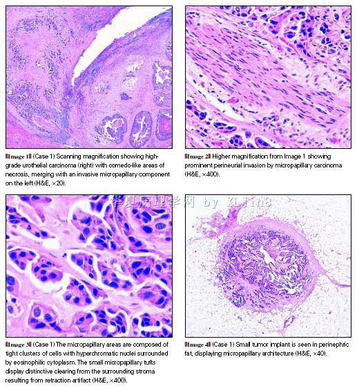
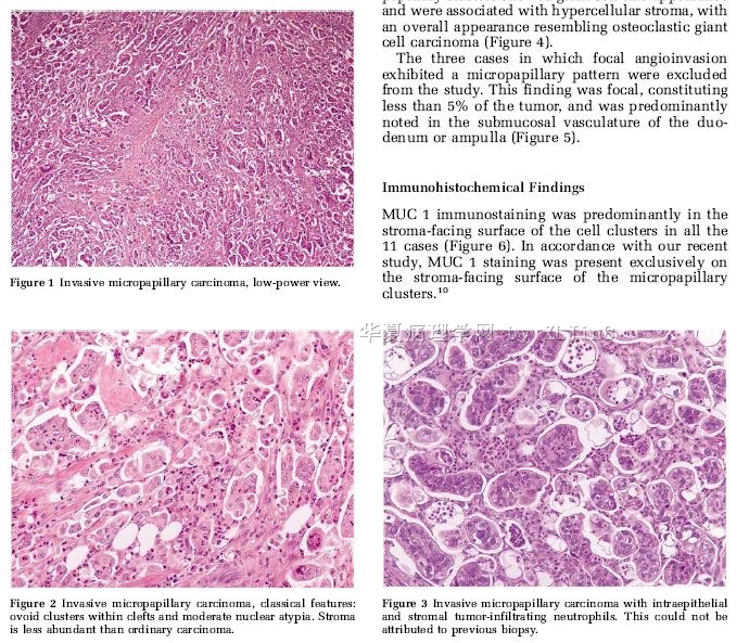
Invasive micropapillary carcinoma has been recently recognized as a rare but distinctive variant of carcinoma in various anatomic sites, including breast, urinary bladder, lung, and major salivary glands(微乳头状癌为癌的一种独特变异型,少见,可发生于不同部位,包括乳腺、膀胱、肺和大唾腺等处). Morphologically, it is characterized by small tight clusters of neoplastic cells floating in clear spaces resembling lymphatic channels. Most often this growth pattern is mixed with a variable component of conventional carcinoma or other variants(形态特征表现为:紧密排列的肿瘤细胞簇团嵌在透亮的腔隙中,就像是漂浮在淋巴管内一样。该长相常多与不同类型的癌成分混合,可以混合普通类型的癌,也可以是与其它变异型的癌成分相混合). In addition to a unique morphology, tumors with invasive micropapillary growth share a high propensity for lymphovascular invasion and lymph node metastases. Patients have typically high-stage disease at presentation and a poor clinical outcome compared with that of patients with conventional carcinoma arising in the same organ site(与相同器官部位发生的传统类型的癌进行比较,除了独特的形态之外,其微乳头生长方式在临床上表现为淋巴管侵袭性强,淋巴结转移率高,患者的临床分期高,预后差). In this article the author reviews the available literature on tumors displaying a micropapillary component.
PMID: 15505530 [PubMed - indexed for MEDLINE]
- 王军臣
-
本帖最后由 于 2011-01-25 13:05:00 编辑
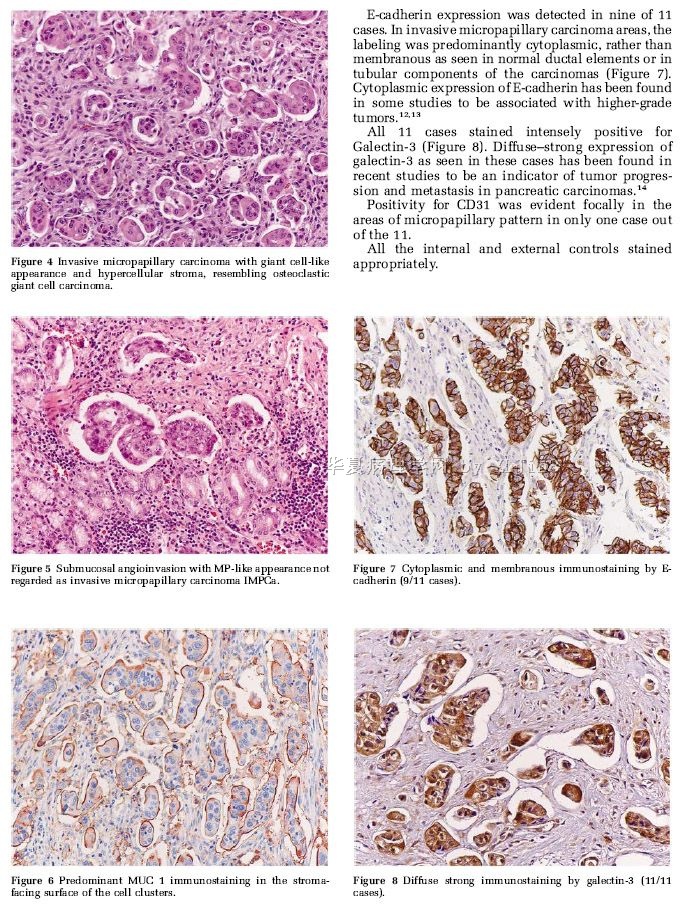
应用抗体配套IHC检测,可以鉴别转移性微乳头状癌的原发部位,除了上述部位外,也有卵巢原发的。
LotanTL,Ye H,MelamedJ,et al.Immunohistochemical panel to identify the primary site of invasive micropapillary carcinoma.Am J Surg Pathol.2009Jul;33(7):1037-41.Invasive micropapillary carcinoma (IMC) is generally an aggressive
morphologic variant that has been described in the bladder, lung, breast,
salivary gland, gastrointestinal tract, and ovary. Given the morphologic
similarities between IMCs arising from different organ systems and the high
propensity of this histologic subtype for lymphatic metastasis, it may be
necessary to use immunohistochemical (IHC) markers to determine the primary
site of an IMC. Few studies have compared the IHC profiles of IMCs originating
from different sites. We tested a panel of 11 IHC markers for their ability to
distinguish urothelial, lung, breast, and ovarian IMC using a tissue microarray
constructed with primary tumor tissue from 47 patients with IMC (13 bladder, 6
lung, 16 breast, and 12 ovarian). For each tumor, correct classification as IMC
was verified by reverse polarity MUC1 expression. We found that immunostaining
for uroplakin, CK20, TTF-1, estrogen receptor (ER), WT-1 and/or PAX8, and
mammaglobin was the best panel for determining the most likely primary site of
IMC. The best markers to identify urothelial IMC were uroplakin and CK20,
whereas p63, high molecular weight cytokeratin, and thrombomodulin were less
sensitive and specific. Lung IMC was uniformly TTF-1 positive. Breast IMC was
ER positive, mammaglobin positive, and PAX8/WT-1 negative, while ovarian IMC was
ER positive, mammaglobin negative, and PAX8/WT-1 positive. In the metastatic
setting, or when IMC occurs without an associated in situ or conventional
carcinoma component, staining for uroplakin, CK20, TTF-1, ER and WT-1, and/or
PAX8, and mammaglobin is the best panel for accurately classifying the likely
primary site of IMC.
- 王军臣
-
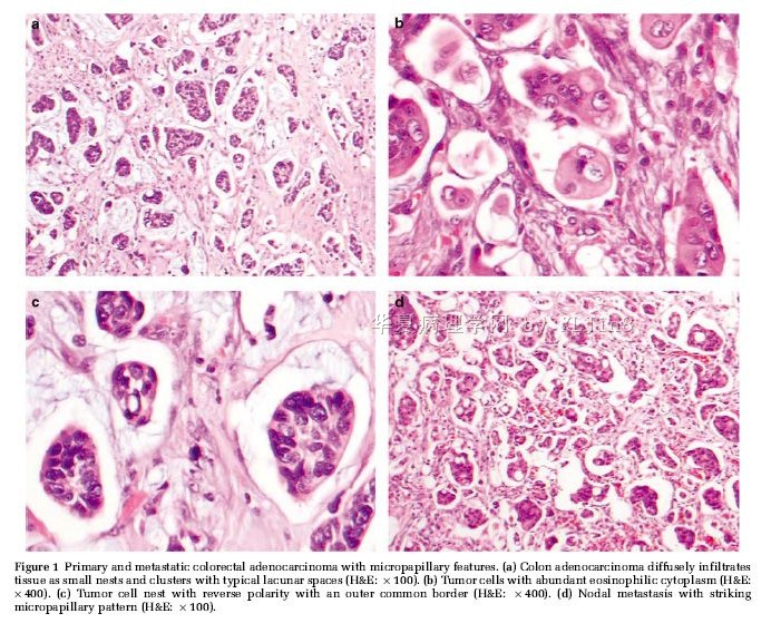
微乳头状癌也可发生于大肠:
Med Mol Morphol. 2007 Dec;40(4):226-30. Epub 2007 Dec 21.
Invasive micropapillary carcinoma of the colon: an immunohistochemical study.
Kuroda N, Oonishi K, Ohara M, Hirouchi T, Mizuno K, Hayashi Y, Lee GH.
Departments of Diagnostic Pathology, Kochi Red Cross Hospital, 2-13-51 Shin-honmachi, Kochi City, Kochi, Japan. nkurodakrch@yahoo.co.jp
Abstract
Invasive micropapillary carcinoma has recently been reported in various anatomic sites. In this article, we report a case of micropapillary carcinoma of the sigmoid colon. A 70-year-old Japanese woman presented with bloody stool for 2 months. Detailed examination disclosed ulcerative and localized tumor in the sigmoid colon. Histological examination of the colon tumor showed a combination of conventional adenocarcinoma (60%) and micropapillary carcinoma (40%). Immunohistochemically, micropapillary carcinoma cells were positive for cytokeratin (CK) 20, carcinoembryonic antigen, and CA125, but negative for CK7, thyroid transcription factor-1, surfactant apoprotein A, estrogen receptor, and progesterone receptor. Additionally, the immunohistochemistry of epithelial membrane antigen revealed reverse polarity of neoplastic cells. Results of conventional adenocarcinoma were basically identical to those of micropapillary carcinoma. In the stroma of both conventional adenocarcinoma and micropapillary carcinoma, many myofibroblasts were present and CD34-positive stromal cells were absent. Finally, we report the fourth case of micropapillary carcinoma arising in the colon. Immunohistochemical results of CK7(-)/CK20(+) strongly suggest the colon as a primary site of micropapillary carcinoma. Additionally, micropapillary carcinoma of the colon may cause a similar stromal reaction to conventional adenocarcinoma of the colon.
- 王军臣
| 以下是引用XLJin8在2011-1-22 17:10:00的发言:
近年来病理医生对癌组织中出现的“微乳头状结构 (micropapillary pattern)”倍加关注,例如肺腺癌、尿路上皮癌、结肠癌、乳腺癌等都有较多文献报道。大多研究认为“微乳头状癌”与预后有关。因此,如何在形态学上把握诊断“微乳头状癌”非常重要,漏了可能导致治疗不足;相反,诊断过了可能导致过度治疗。请感兴趣的网友发表意见。谢谢! 以下为有关病例,请参阅。 http://www.ipathology.cn/forum/forum_display.asp?keyno=342819 http://www.ipathology.cn/forum/forum_display.asp?keyno=278637 http://www.ipathology.cn/forum/forum_display.asp?keyno=342108
请金老师讲课!!!! | ||||||||||||||||||||||||
-
cnlzh20060 离线
- 帖子:224
- 粉蓝豆:58
- 经验:378
- 注册时间:2009-02-27
- 加关注 | 发消息
-
xuchuanjie 离线

- 帖子:4
- 粉蓝豆:81
- 经验:53
- 注册时间:2008-01-09
- 加关注 | 发消息


