| 图片: | |
|---|---|
| 名称: | |
| 描述: | |
- 特殊类型的肾脏肿瘤
| 姓 名: | ××× | 性别: | 男 | 年龄: | 52 |
| 标本名称: | |||||
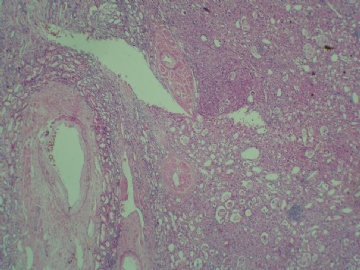
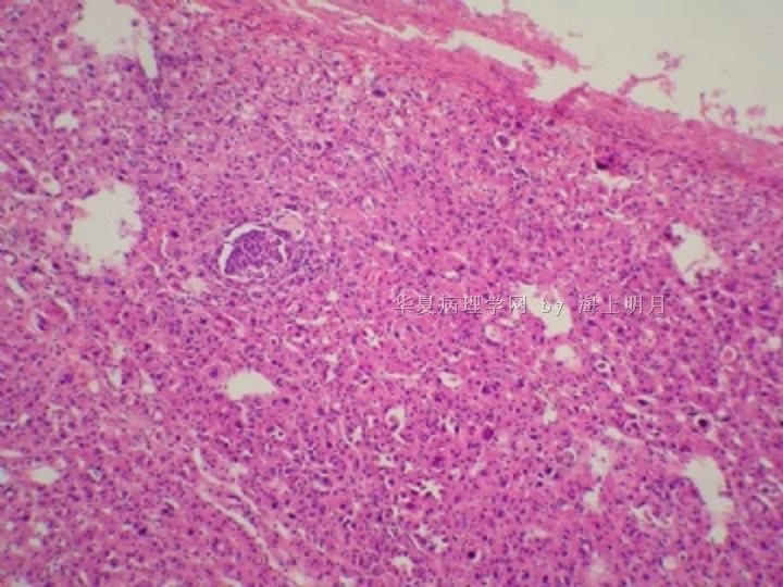
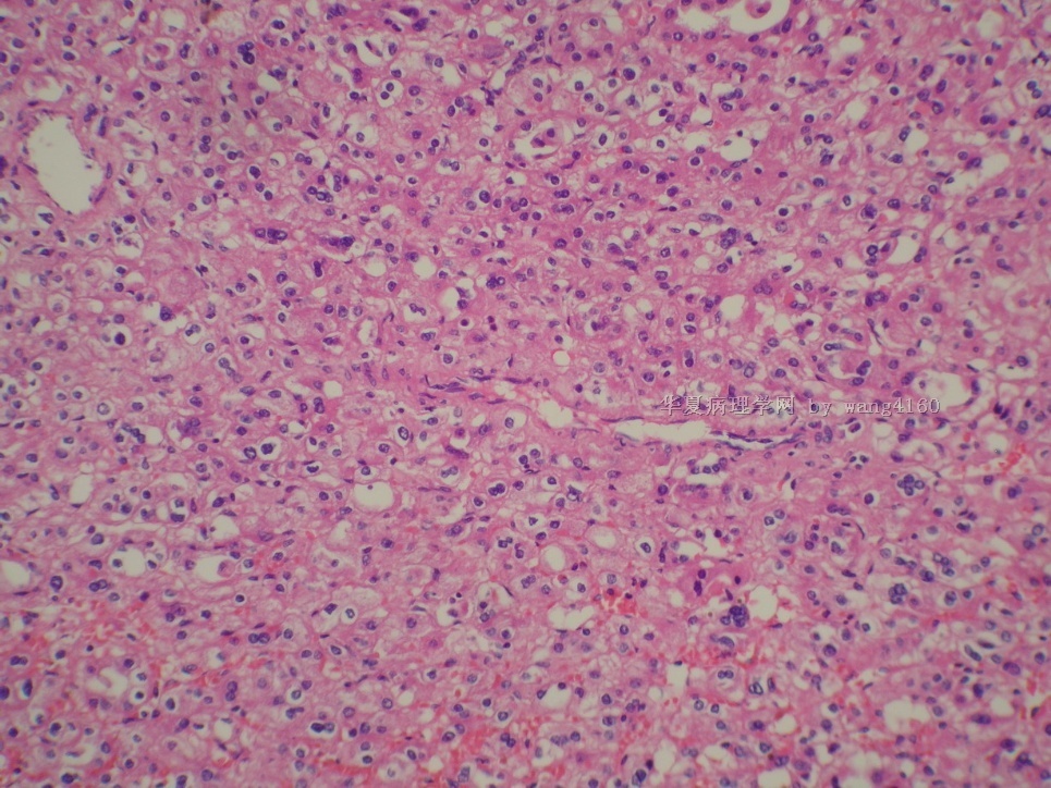
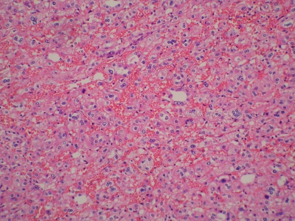
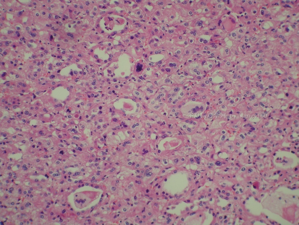
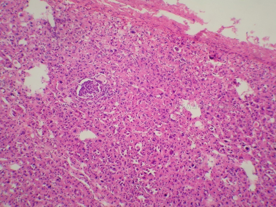
| 简要病史: | 体检发现腹腔肿块,无血尿,尿潴留 | ||||
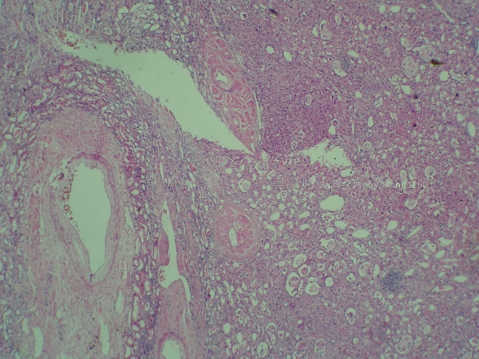
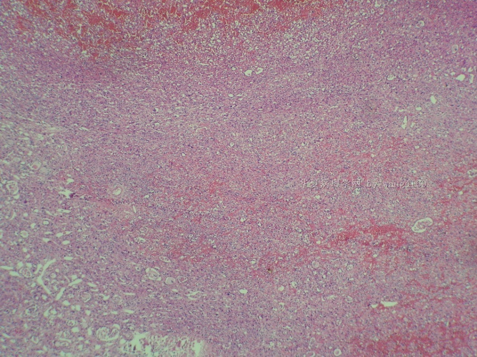
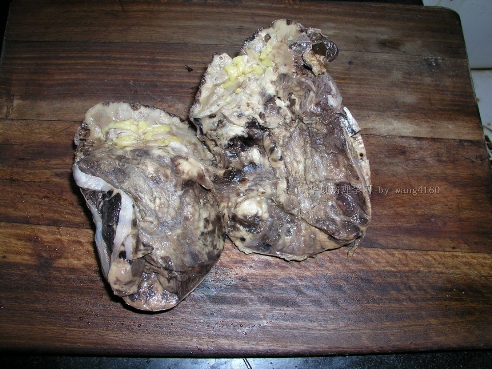
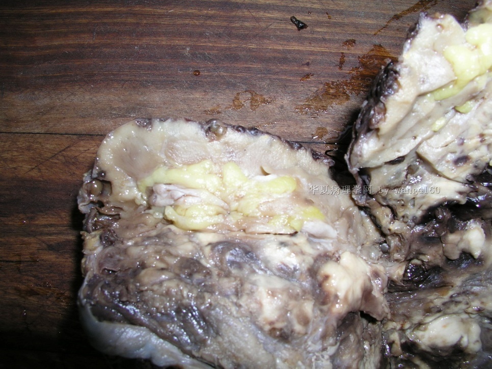
| 肉眼检查: | 术中发现与肾脏、胰腺粘连,大体切面灰白实性,有出血 | ||||
名称:图1
描述:图1
名称:图2
描述:图2
名称:图3
描述:图3
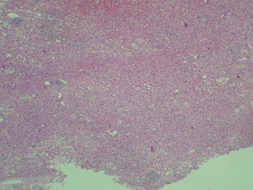
名称:图4
描述:图4
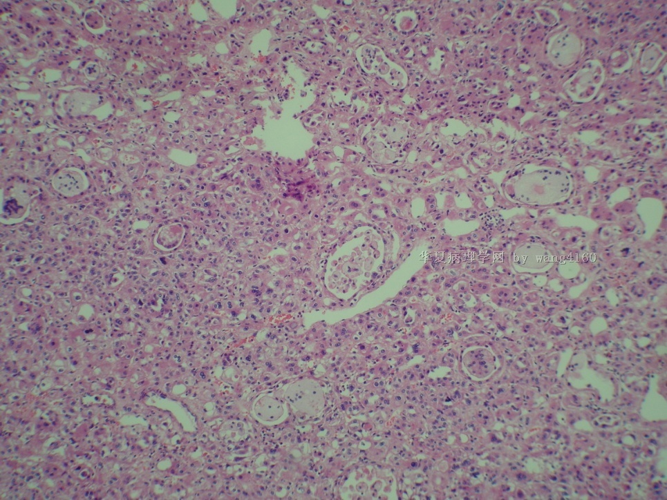
名称:图5
描述:图5
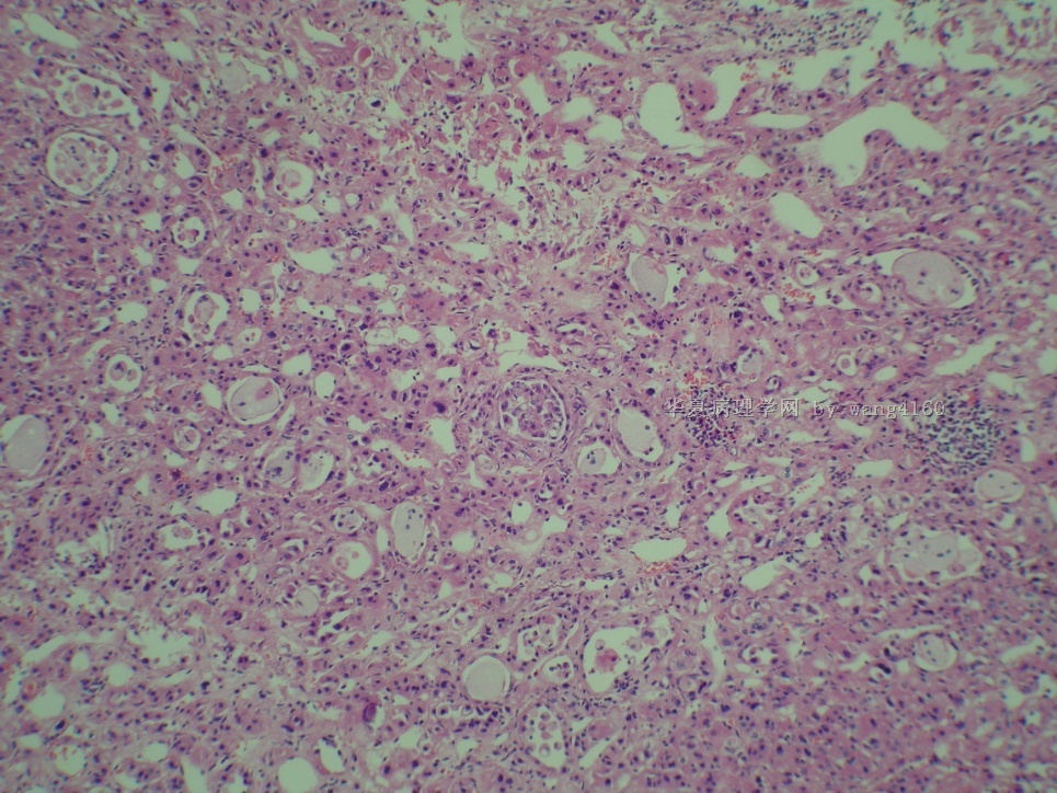
名称:图6
描述:图6
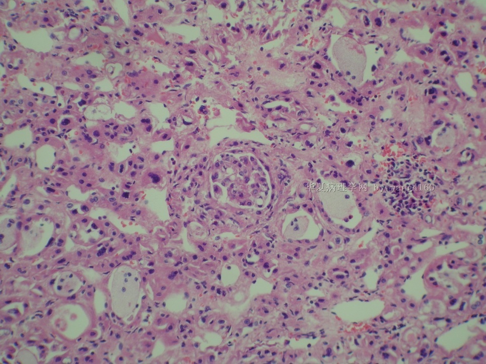
名称:图7
描述:图7
名称:图8
描述:图8
名称:图9
描述:图9
名称:图10
描述:图10
名称:图11
描述:图11
名称:图12
描述:图12
名称:图13
描述:图13
名称:图14
描述:图14
名称:图15
描述:图15
名称:图16
描述:图16
名称:图17
描述:图17
-
本帖最后由 于 2011-01-11 07:38:00 编辑
-
cnlzh20060 离线
- 帖子:224
- 粉蓝豆:58
- 经验:378
- 注册时间:2009-02-27
- 加关注 | 发消息
-
本帖最后由 于 2011-01-10 09:11:00 编辑
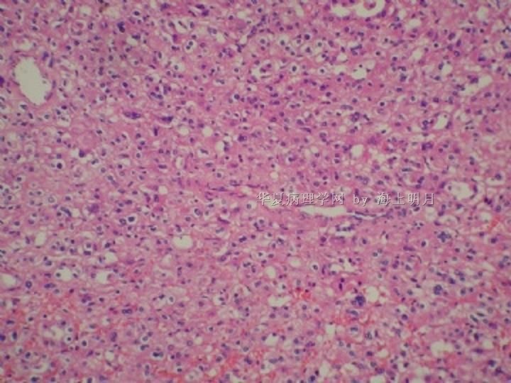
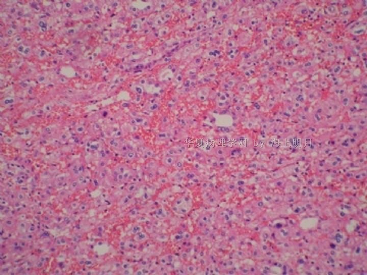
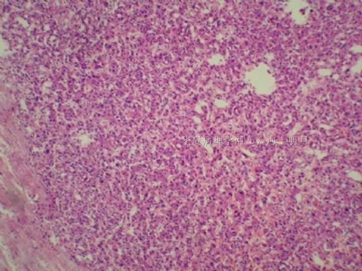
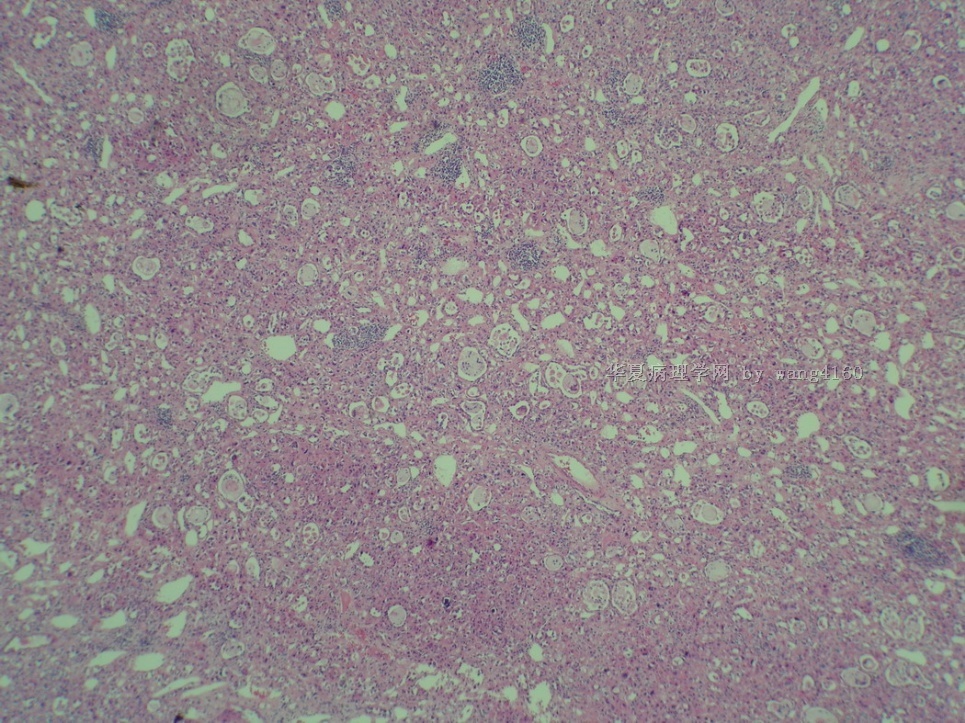
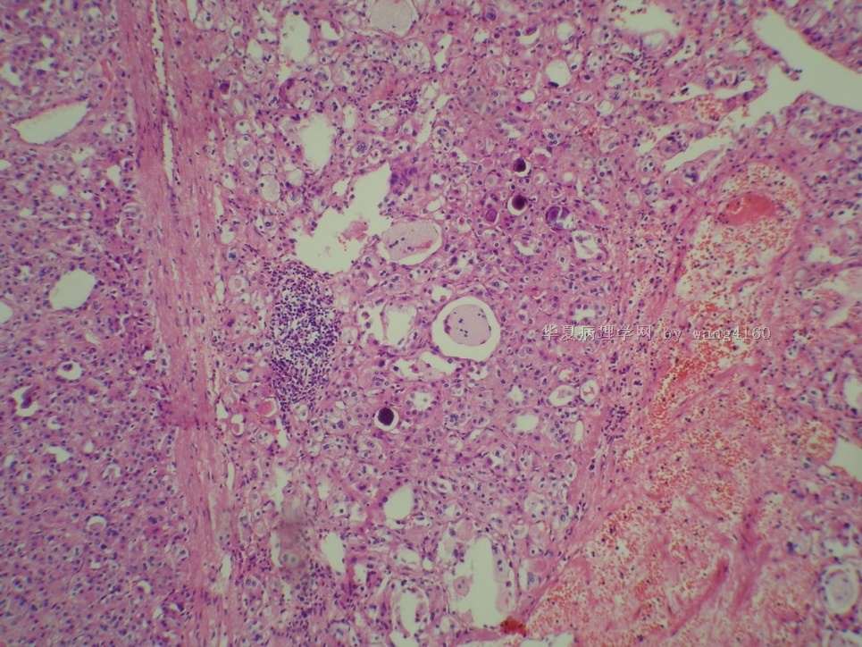
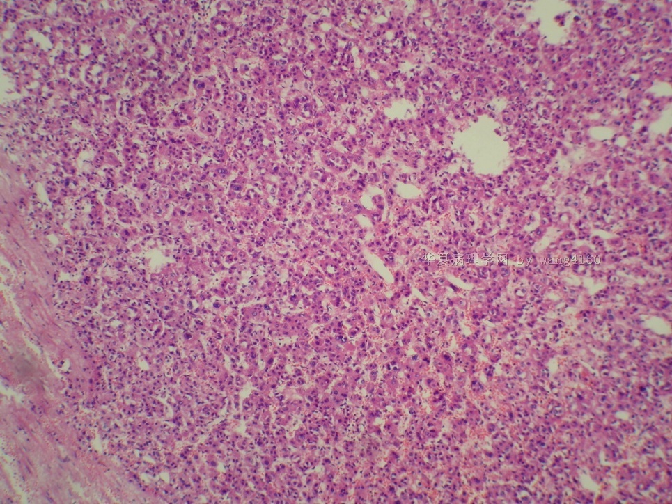
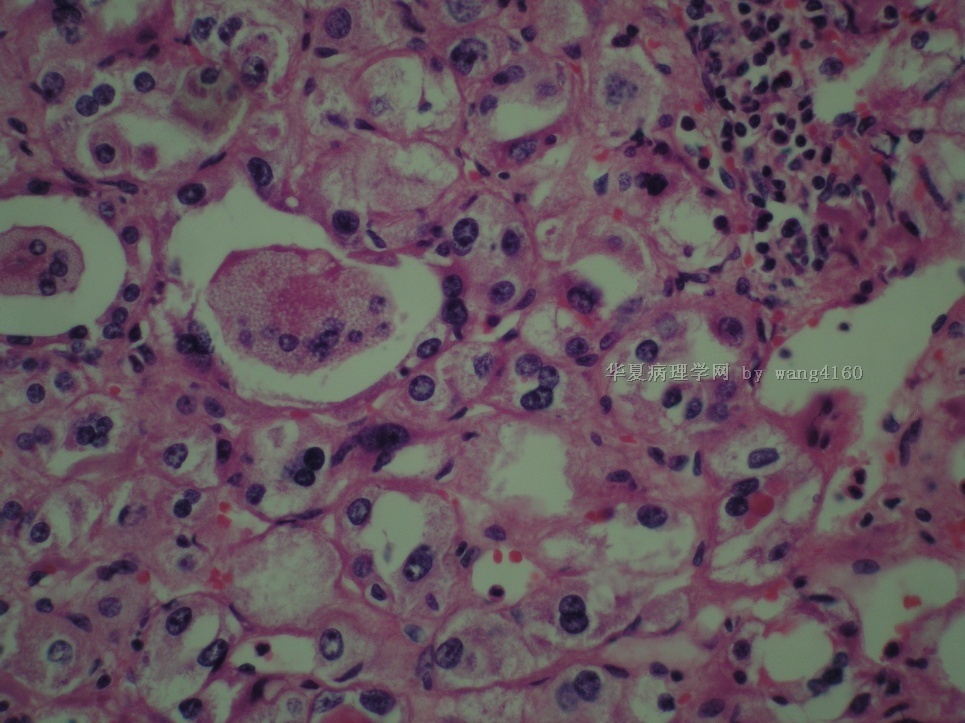
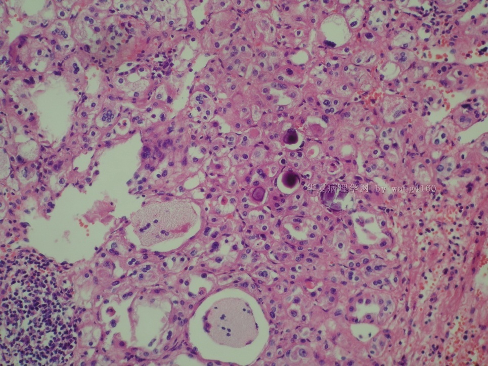
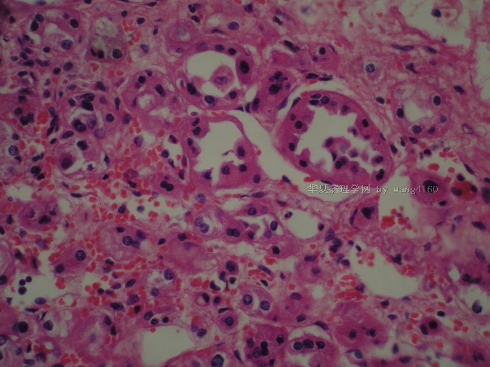
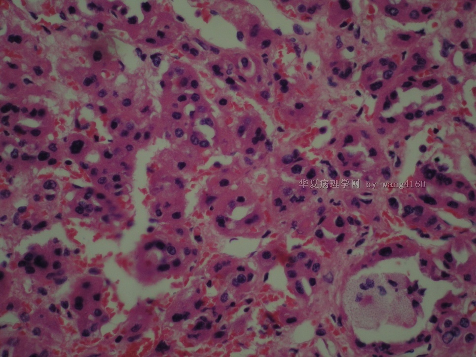
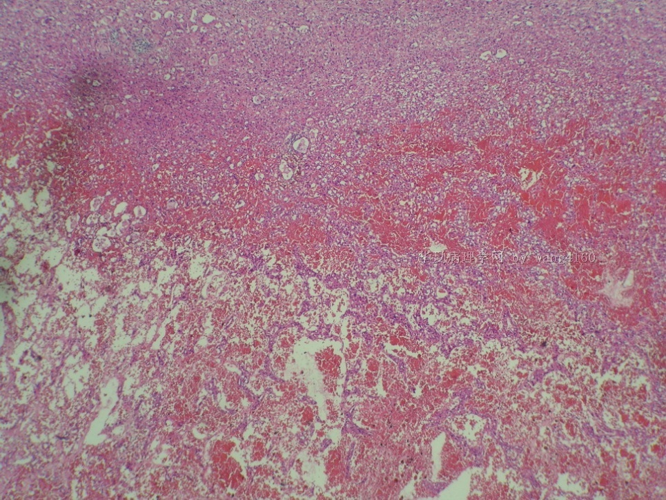
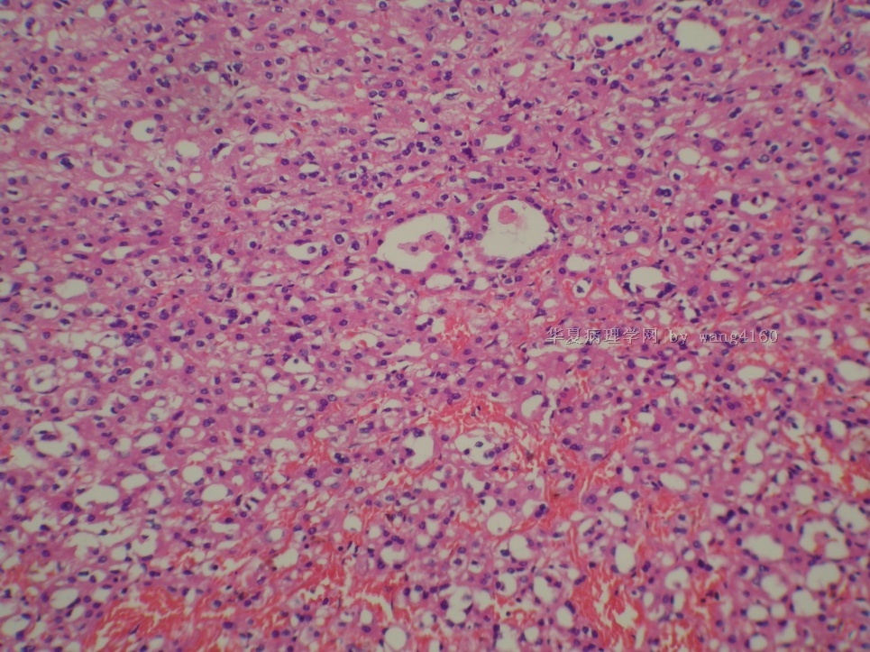
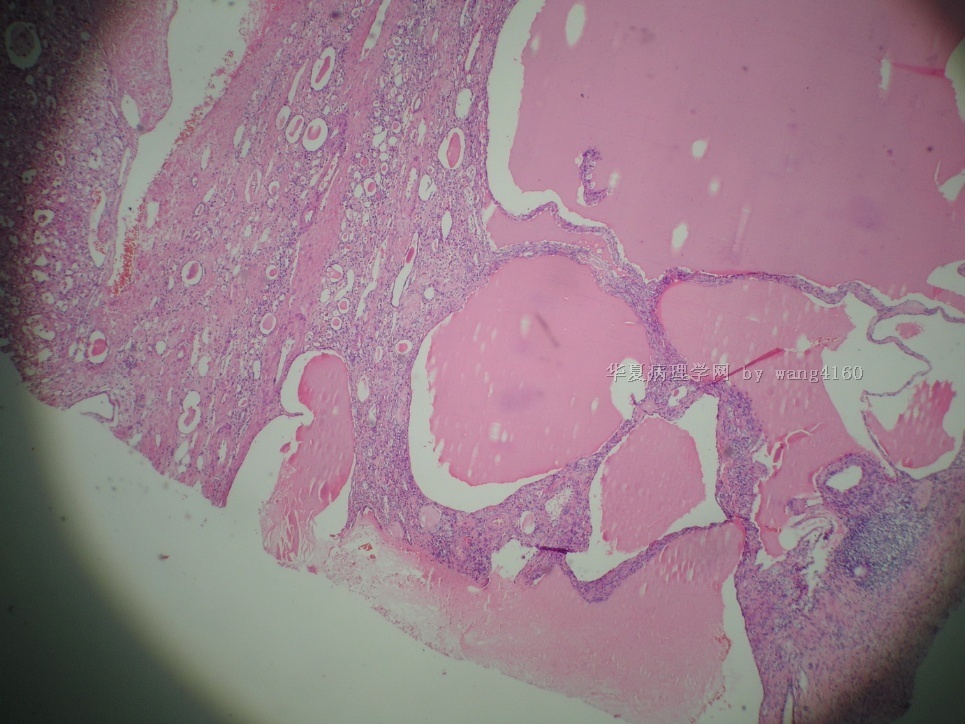
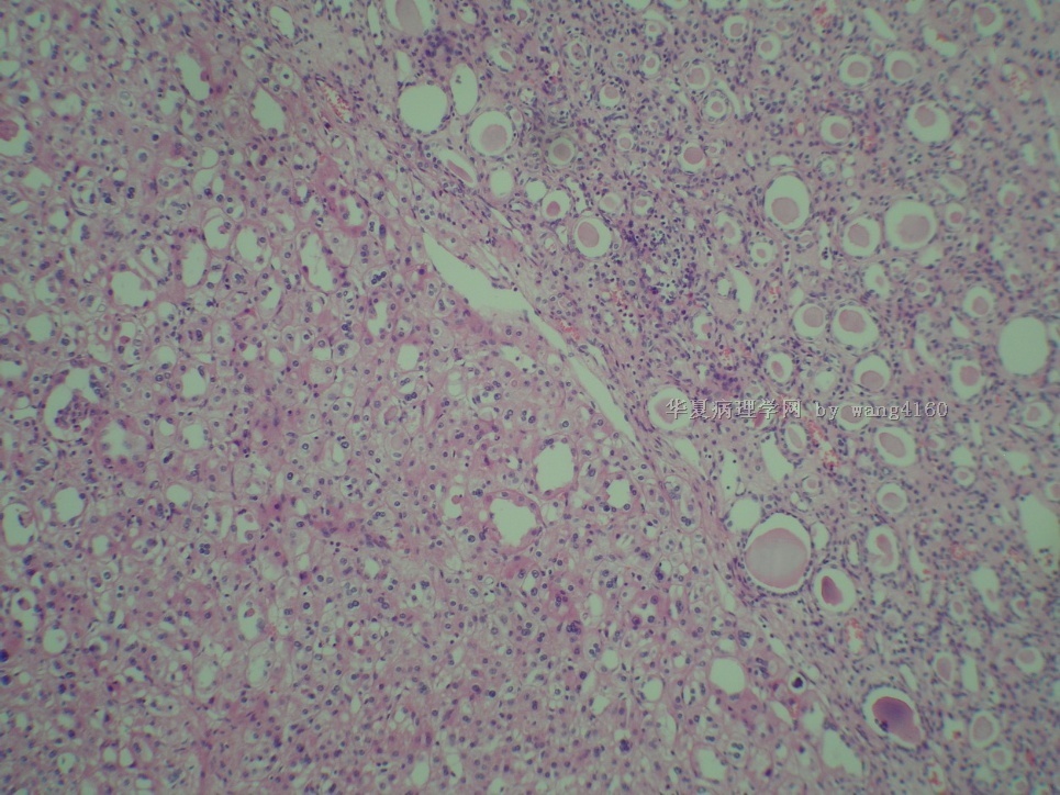
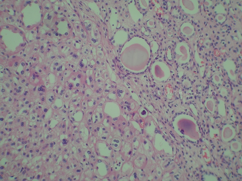
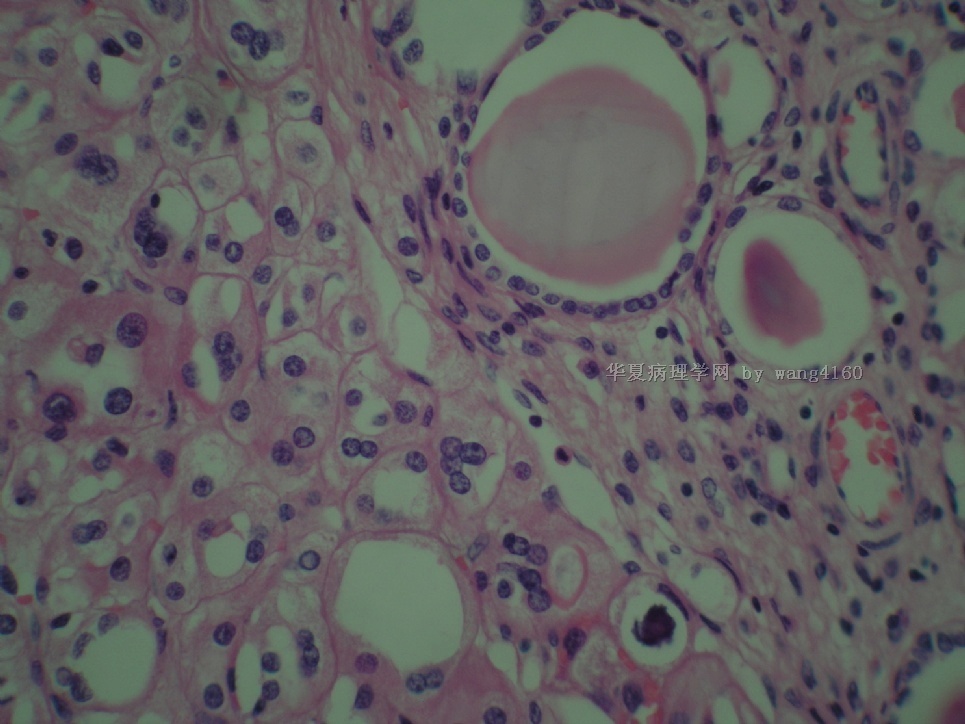
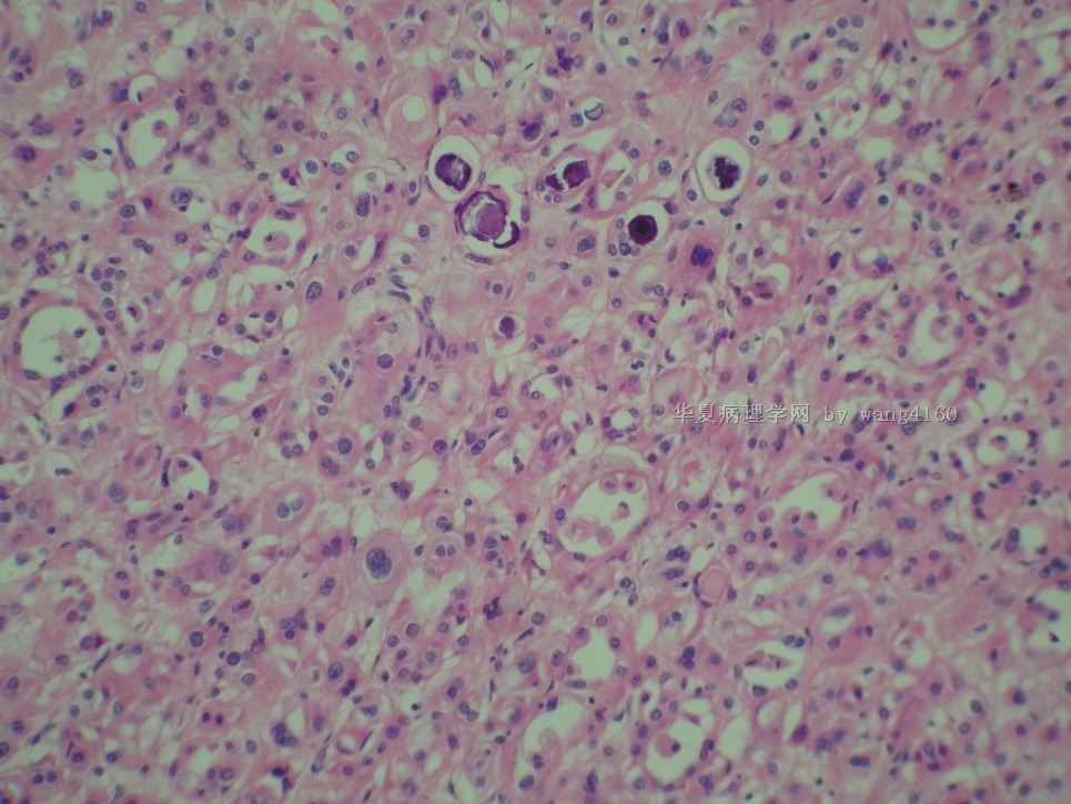
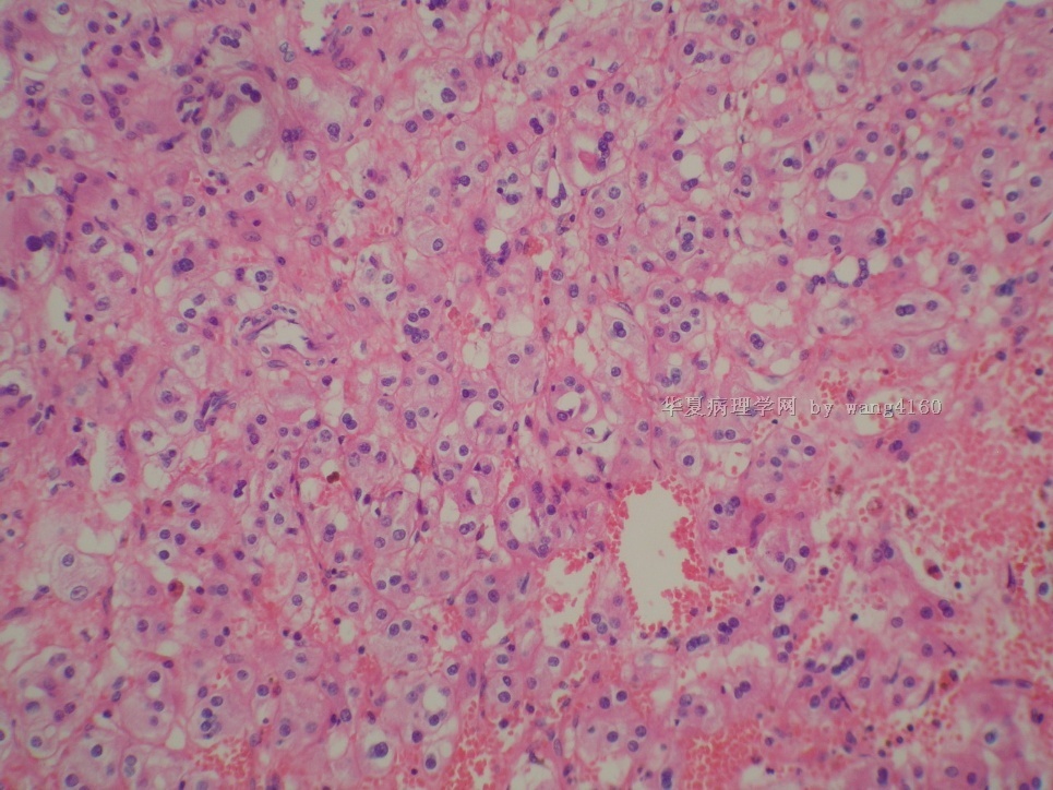
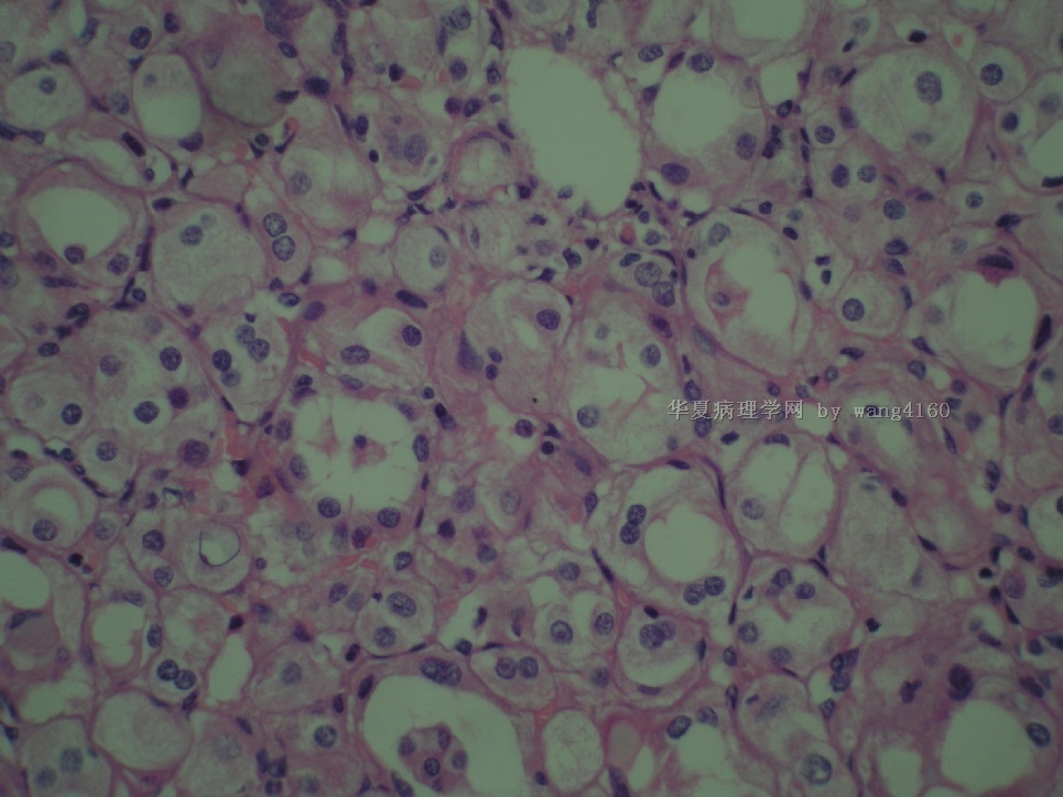
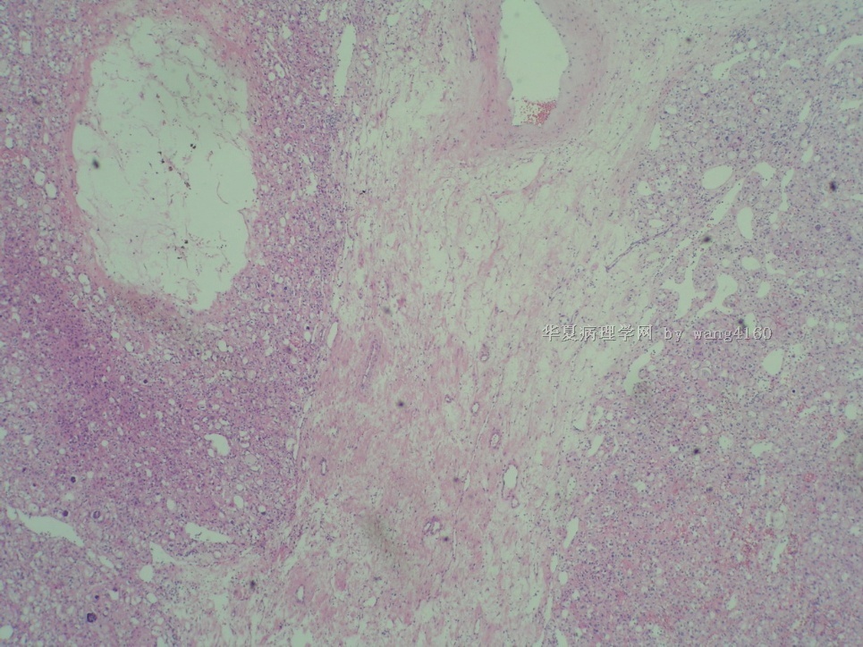
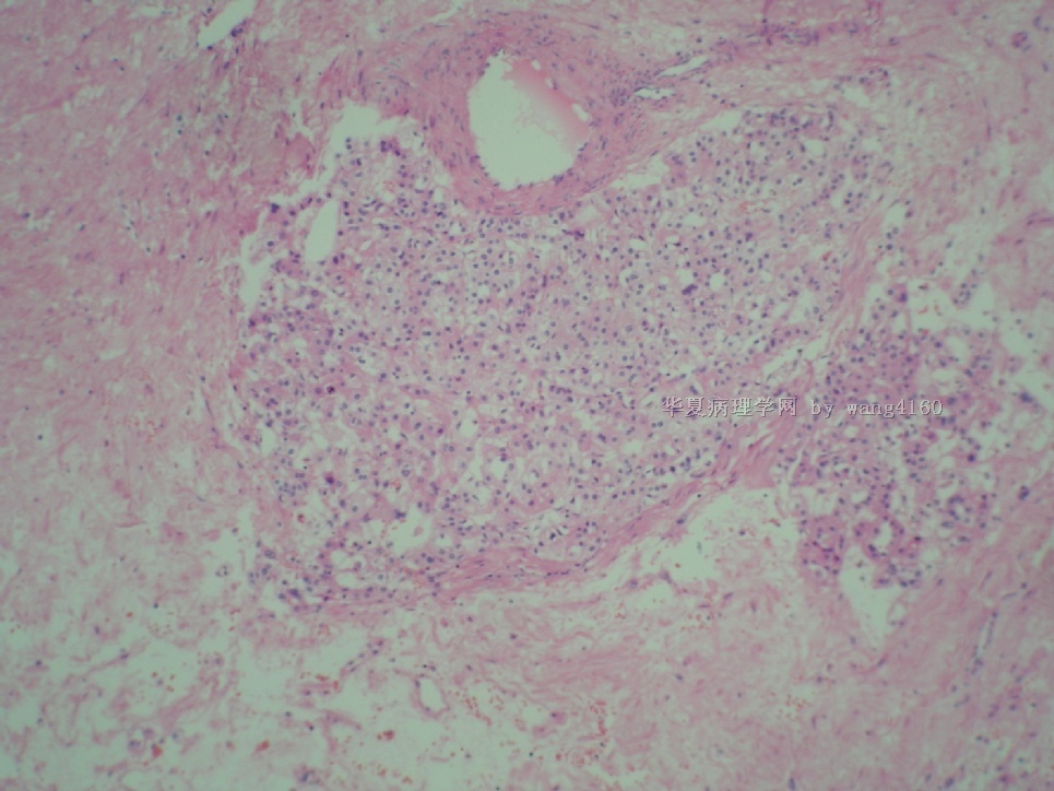
再上传一些图片和大体照片,科室主要意见也是肾细胞癌,但是实在没见过这样的,管状结构发育如此明显,同时Villin和CD10又阴性(内对照肾小管阳性)!
名称:图1
描述:图1
名称:图2
描述:图2
名称:图3
描述:图3
名称:图4
描述:图4
名称:图5
描述:图5
名称:图6
描述:图6
名称:图7
描述:图7
名称:图8
描述:图8
名称:图9
描述:图9
名称:图10
描述:图10
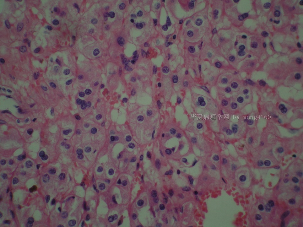
名称:图11
描述:图11
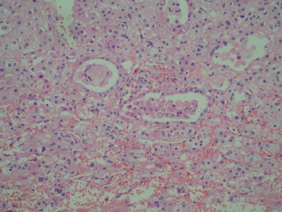
名称:图12
描述:图12
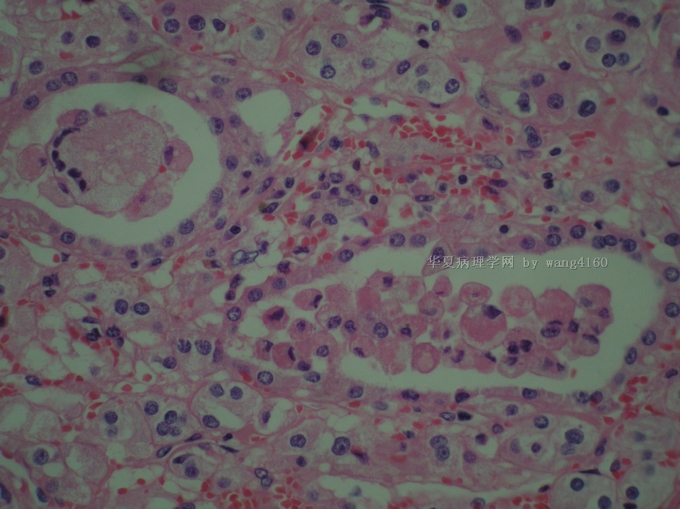
名称:图13
描述:图13
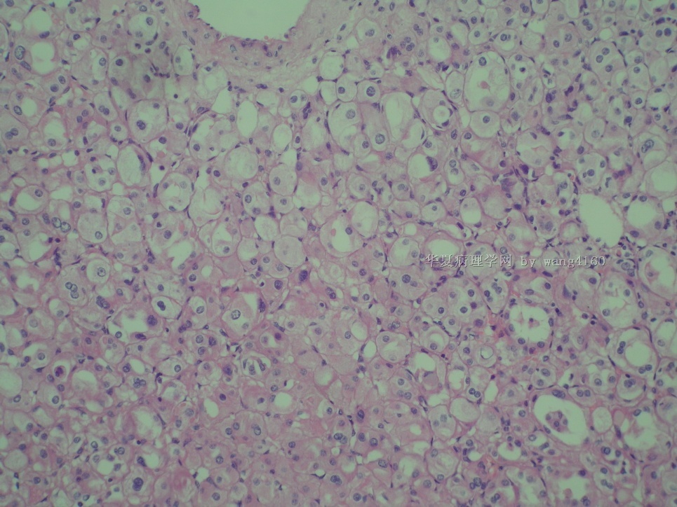
名称:图14
描述:图14
名称:图15
描述:图15
名称:图16
描述:图16
名称:图17
描述:图17

名称:图18
描述:图18
-
本帖最后由 于 2011-01-11 07:46:00 编辑
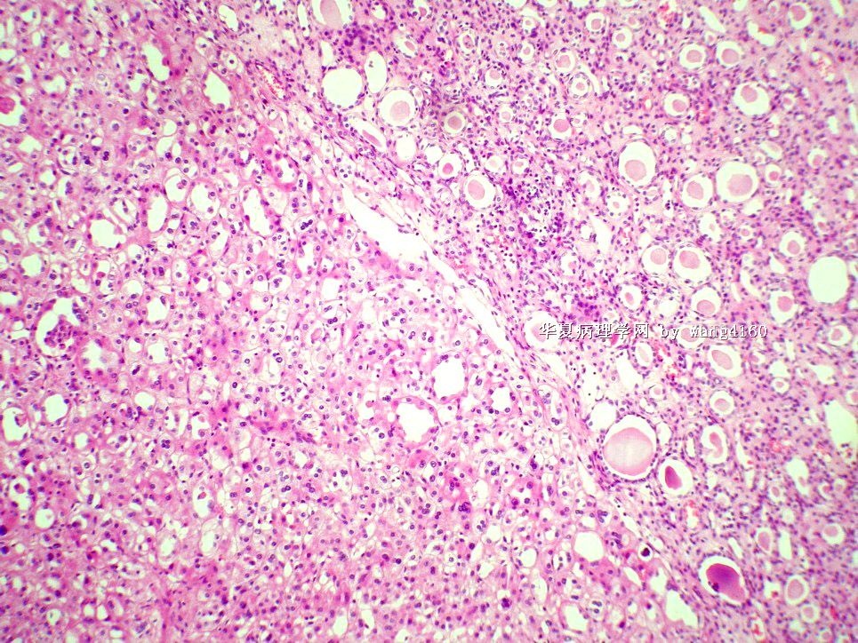
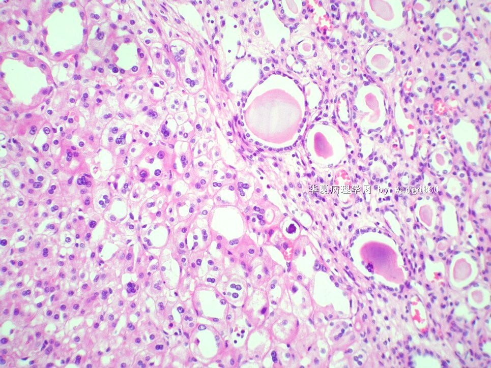
形态学非常特殊的成人肾脏肿瘤。有些区域很像甲状腺滤泡,因此,是否就是晚近报道的肾癌新类型-肾脏甲状腺滤泡样癌(Thyroid-like follicular carcinoma of the kidney)?世界上仅10多例报道。此例很可能为中国首例TLFCK。
希望能多做些工作, 尽快发表。
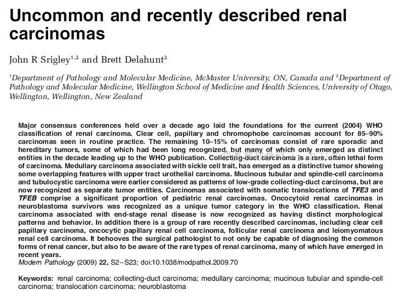
图1-2引用文献,图3-4为本病例。
感谢楼主分享如此好的罕见病例!

名称:图1
描述:图1

名称:图2
描述:图2
名称:图3
描述:图3
名称:图4
描述:图4
名称:图5
描述:图5

名称:图6
描述:图6

- xljin8
-
本帖最后由 于 2011-01-11 07:50:00 编辑
参考文献:
1. Hum Pathol. 2011 Jan;42(1):146-50. Epub 2010 Oct 23.
Thyroid-like follicular carcinoma of the kidney with metastases to the lungs and retroperitoneal lymph nodes.
Dhillon J, Tannir NM, Matin SF, Tamboli P, Czerniak BA, Guo CC.
Department of Pathology, The University of Texas MD Anderson Cancer Center, Houston, TX 77030-4009, USA.
Thyroid-like follicular carcinoma of the kidney is an extremely rare variant of renal cell carcinoma. Most previously reported cases presented as incidental small tumors confined to the kidney. Here we report a unique case in which the patient presented with flank pain and hematuria. Imaging studies demonstrated a large tumor in the right kidney and metastases to the lungs and retroperitoneal lymph nodes. Both the renal tumor and the sampled lung metastasis were composed almost entirely of follicles with dense, colloid-like material resembling thyroid follicular carcinoma. However, no lesion was found in the thyroid gland; and the patient's thyroid function test results were normal. The tumor cells were immunoreactive for PAX2 and PAX8 but lacked reactivity for thyroglobulin and thyroid transcription factor-1. To our knowledge, this is the first case of thyroid-like follicular carcinoma of the kidney to be initially associated with marked symptoms and widespread metastases, providing evidence that this rare variant of renal cell carcinoma can be clinically aggressive.
2. Am J Surg Pathol. 2009 Mar;33(3):393-400.
Primary thyroid-like follicular carcinoma of the kidney: report of 6 cases of a histologically distinctive adult renal epithelial neoplasm.
Amin MB, Gupta R, Ondrej H, McKenney JK, Michal M, Young AN, Paner GP, Junker K, Epstein JI.
Department of Pathology, Cedars-Sinai Medical Center, Los Angeles, CA 90048, USA.aminm@cshs.org
Thyroidization of kidney reminiscent of thyroid follicles with accumulation of inspissated colloid-like material in renal tubules is a hallmark of chronic pyelonephritis. We identified 6 tumors in the kidney, distinct from currently known subtypes of renal cell carcinoma, with a striking histology that closely mimicked well-differentiated thyroid follicular neoplasms and raised the possibility of metastatic follicular thyroid carcinoma. Three occurred in males and 3 in females with an age range of 29 to 83 years and size range from 1.9 to 4 cm. All tumors were encapsulated and exclusively demonstrated follicular architecture comprising of microfollicles and macrofollicles containing inspissated colloid-like material. A minor component of small tightly packed follicles devoid of secretions was also noted. The follicles were lined by cells
with moderate amphophilic to eosinophilic cytoplasm with round nuclei and occasional prominent nucleoli. The tumors were nonimmunoreactive with thyroglobulin and thyroid transcription factor 1 and for markers contemporarily used for renal differentiation. The tumors had a gene expression profile distinct from clear cell and chromophobe renal cell carcinoma. Comparative genetic hybridization failed to reveal cytogenetic alterations. Mean follow-up of 47.3 months (range: 7 to 84 mo) showed that 5 patients had no evidence of disease and 1 developed a metastasis to the renal hilar lymph nodes in which the follicular architecture with colloid was retained. Thyroid-like follicular renal cell carcinoma represents a unique histologic subtype of renal cell carcinoma of low malignant potential and its primary importance is to distinguish it from metastatic carcinoma from the thyroid. 3. Virchows Arch. 2008 Jan;452(1):91-5. Epub 2007 Aug 18. Thyroid follicular carcinoma-like renal tumor: a case report with morphologic,immunophenotypic, cytogenetic, and scintigraphic studies. Sterlacci W, Verdorfer I, Gabriel M, Mikuz G. Department of Pathology, Medical University of Innsbruck, Muellerstr 44,Innsbruck, Austria. william.sterlacci@i-med.ac.at Erratum in:Virchows Arch. 2008 Apr;452(4):471. William, Sterlacci [corrected to Sterlacci,William]; Irmgard, Verdorfer [corrected to Verdorfer, Irmgard]; Michael, Gabriel [corrected to Gabriel, Michael]; Gregor, Mikuz [corrected to Mikuz, Gregor]. Comment in:Virchows Arch. 2009 Jun;454(6):717-8. In this report, a rare renal tumor that morphologically resembles a thyroid follicular carcinoma is described. To date, this subtype has not been integrated into a known form of renal carcinoma. A 29-year-old female patient without relevant family or social history underwent nephrectomy because of a renal tumor measuring 5 cm by the largest diameter. The macroscopically white-yellow tumor showed follicular structures with abundant eosinophilic colloidal material and focal papillary differentiation by light microscopy. Immunohistochemically, the tumor cells stained positively for cytokeratin (CK-7, CK-20, CAM 5.2) and vimentin. CD-10, CD-117, thyroid transcription factor-1, and thyreoglobulin remained completely negative. Chromosomal losses of 1, 3, 7, 9p21, 12, 17, and X were detected by fluorescence in situ hybridization. Scintigraphs showed an inconspicuous thyroid gland and no extrathyroidal pathological accumulations,making metastatic spread to the kidney highly unlikely. To our knowledge, this is the second fully documented case of a thyroid follicular carcinoma-like renal tumor. This uncommon variant is important to keep in mind to prevent unnecessary or inappropriate treatment. 4. Am J Surg Pathol. 2006 Mar;30(3):411-5. Thyroid follicular carcinoma-like tumor of kidney: a case report with morphologic, immunohistochemical, and genetic analysis. Jung SJ, Chung JI, Park SH, Ayala AG, Ro JY. Department of Pathology, Inje University College of Medicine, Busan, Korea.soojinmd@hanmail.net We present an unusual renal tumor, which has not been classified under a known subtype of renal cell carcinoma (RCC) and characteristically shows similar histology to thyroid follicular carcinoma. The patient was a 32-year-old asymptomatic woman who was found to have a kidney mass during her annual physical examination. She had no lesions in the thyroid during physical and ultrasound examinations, and there was no abnormal thyroid function test. Neither mediastinal nor ovarian abnormalities were observed. The resected kidney showed a well-defined nodular tumor measuring 11.8x8.0x8.0 cm. The mass was protruding into the pelvic cavity with areas of yellowish geographic necrosis. Histologically, the tumor showed follicular architectures with inspissated colloid-like material in their lumina. No conventional (clear cell) RCC or any other known subtypes of RCC component was observed. Immunohistochemically, the tumor cells showed intensive staining for cytokeratin (CK) cocktail AE1/AE3 and CD10 and were not reactive to thyroid transcription factor-1 and thyroglobulin. The staining of CK35betaH11 and vimentin revealed focal cytoplasmic reaction. The tumor cells were completely negative for CK7, CK19, CK20, CK34betaE12, carcinoembryonic antigen, epithelial membrane antigen, and CD15. Chromosomal gains of 7q36, 8q24, 12, 16, 17p11-q11, 17q24, 19q, 20q13, 21q22.3, and Xp and losses of 1p36, 3, and 9q21-33 were detected by comparative genomic hybridization. These findings are dissimilar to previously classified renal neoplasm. Only a report that included three cases of primary thyroid-like renal tumor has been described in the abstract form. However, there is no fully documented case on this unusual form of RCC, which morphologically resembles that of thyroid follicular carcinoma. Herein, we present a new case of thyroid follicular carcinoma-like tumor of the kidney with a chromosomal study and review of the literature.

- xljin8
-
本帖最后由 于 2011-01-12 15:08:00 编辑
请见目前发表的英文相关参考文献:
Results: 5

- 王军臣

