| 图片: | |
|---|---|
| 名称: | |
| 描述: | |
- 女/17岁 左大脑颞叶结节状囊内病变(2010上海-大阪-墨尔本读片会) Dr. Tony Thomas提供
-
zhaoyan2006 离线
- 帖子:139
- 粉蓝豆:42
- 经验:301
- 注册时间:2009-02-23
- 加关注 | 发消息
-
本帖最后由 于 2010-12-15 06:53:00 编辑
常可表现为囊性病变的脑神经细胞和胶质细胞的肿瘤包括:
1)毛细胞星形细胞瘤(pilocytic astrocytoma);
2)多形性黄色星形细胞瘤(pleomorphic xanthoastrocytoma);
3)节细胞胶质瘤(ganglioglioma);
4)促纤维增生性婴儿节细胞胶质瘤(desmoplastic infantile ganglioglioma);
5)脑室内神经细胞瘤( extraventricular neurocytic neoplasm);
6)室管膜瘤(ependymoma);
7)星形母细胞瘤( astroblastoma);
此病例确实非常难(上海华山医院汪寅教授也认为非常难,从未见到过类型的脑肿瘤),世界文献中至今尚未报道此变型(variant)。
非常有挑战性的病例!欢迎大家参加讨论。

- xljin8
| 以下是引用xljin8在2010-12-15 5:58:00的发言:
常可表现为囊性病变的脑神经细胞和胶质细胞的肿瘤包括: 1)毛细胞星形细胞瘤(pilocytic astrocytoma); 2)多形性黄色星形细胞瘤(pleomorphic xanthoastrocytoma); 3)节细胞胶质瘤(ganglioglioma); 4)促纤维增生性婴儿节细胞胶质瘤(desmoplastic infantile ganglioglioma); 5)脑室内神经细胞瘤( extraventricular neurocytic neoplasm); 6)室管膜瘤(ependymoma); 7)星形母细胞瘤( astroblastoma);
此病例确实非常难(上海华山医院汪寅教授也认为非常难,从未见到过类型的脑肿瘤),世界文献中至今尚未报道此变型(variant)。 非常有挑战性的病例!欢迎大家参加讨论。 |
“此病例确实非常难(上海华山医院汪寅教授也认为非常难,从未见到过类型的脑肿瘤),世界文献中至今尚未报道此变型(variant)。”看来是不简单,几天了没有人发言,既然已经丢丑了,那就继续丢下去了:
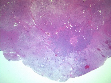
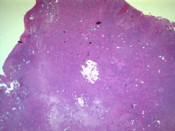
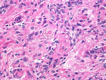
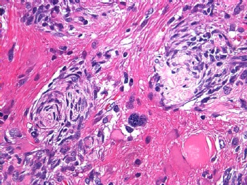
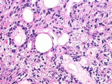
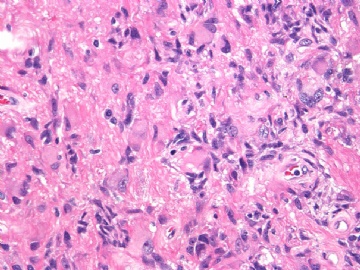
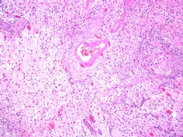
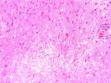
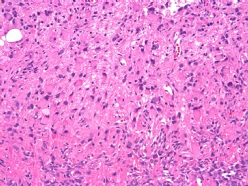
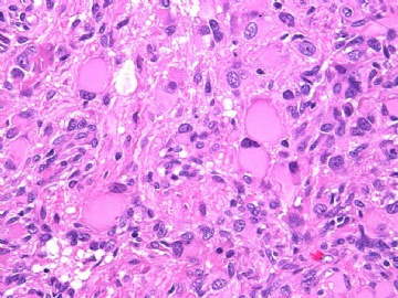
本例组织学特征如下:
17岁颅内(幕上)结节状囊性病变,
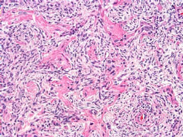
有促纤维增生性的星形胶质细胞
可见Rosenthal纤维,
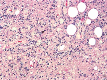
有片状分布的核偏位的胞浆嗜酸性横纹肌样细胞(不像节细胞),
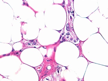
有灶状成熟的脂肪细胞等,
-
lantian0508 离线
- 帖子:1250
- 粉蓝豆:42
- 经验:1495
- 注册时间:2007-08-01
- 加关注 | 发消息
Case comes from
This is challenging case. I have not ever seen any other complicated lesion like this case.
In clinically, a young woman presented with epilepsy. Brain MRI reveals a nodular mass with cystic formation on the cortical surface in left temporal lobe.
Histopathologically, I think this tumor at least showed four histopathological patterns.
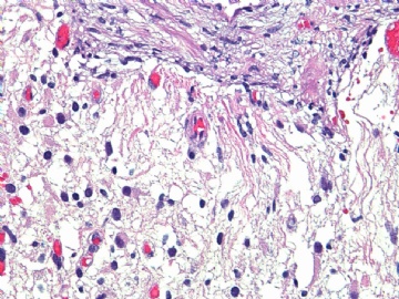
The first is presence of glial background admixed with neuronal components. The tumor cells disclose pleomorphism, pale or lipid-filled cytoplasm. In some areas, I can see some medium-sized mature ganglion-like cells and neuropil-like matrix. However, there are no presences of typical large ganglion cells. Lack of dominant mitotic activity, microvascular proliferation and necrosis.
The second is presence of dominant mesenchymal components on the cortical surface accompany with focal lipomatous metaplasia.
The third is occasionally presence of pseudopapillary structure formations, which shows hyalinized blood vessels surrounded by a single layer of tumor cells.
The fourth is presence of degenerative changes such as Rosenthal fibers and eosinophilic granular bodies. But, there are no dominant lymphocytic infiltrations.
Based on these histopathological findings, I can not make an accurate diagnosis according to current issue of WHO classification tumors of the CNS. Thus, I prefer a diagnosis of pleomorphic xanthoastrocytoma (PXA)-like unclassified low grade glioneuronal tumor with focal mesenchymal differentiation, corresponding to WHO grade II is the most possibility for this case.
The differential diagnosis should be including ganglioglioma and lipoastrocytoma.
By the way
At present, PXA is also considered a mixed type of glioneuronal tumor, but is not pure astrocytic tumor.
