| 图片: | |
|---|---|
| 名称: | |
| 描述: | |
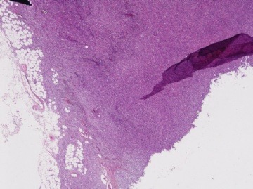
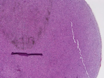
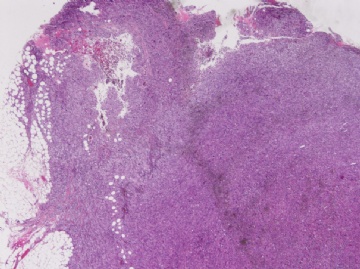
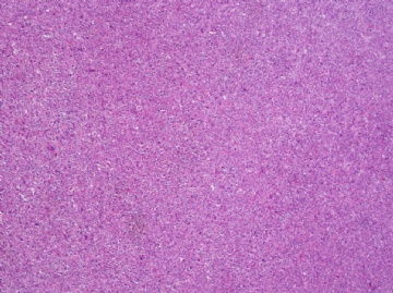
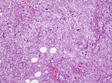
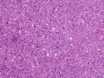
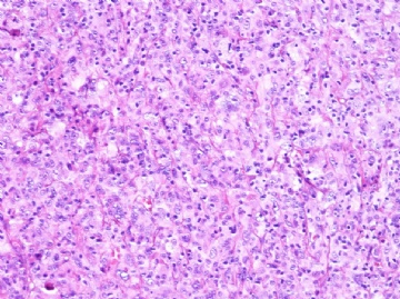
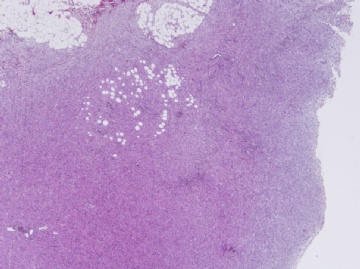
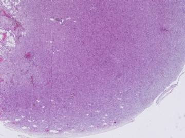
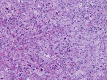
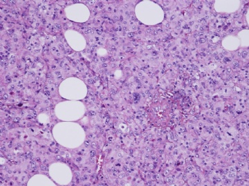
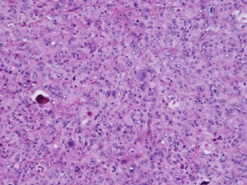
- 后腹膜病例-地坛
-
Epithelioid variant of pleomorphic liposarcoma: a comparative immunohistochemical and ultrastructural analysis of six cases with emphasis on overlapping features with epithelial malignancies.
Department of Pathology, Memorial Sloan-Kettering Cancer Center, New York, New York, USA.
Ultrastruct Pathol. 2002 Sep-Oct;26(5):299-308.
Abstract
Pleomorphic liposarcoma (PL) is the least common subtype of liposarcoma, displaying a lipoblastic, malignant fibrous histiocytoma (MFH)-like and, less frequently, an epithelioid growth pattern. The epithelioid morphology in PL is still underrecognized and may closely simulate other epithelial neoplasms, mainly adrenal cortical carcinoma (ACC). No electron microscopic (EM) studies of the epithelioid variant of PL have been previously described, nor have there been studies of its immunoreactivity with A103 or alpha-inhibin. The purpose of this study is to analyze the histological, immunohistochemical, and EM features of epithelioid PL in an attempt to better explore the distinction from their epithelial mimickers, such as ACC. A panel of 5 antibodies was studied, including A103, alpha-inhibin, smooth muscle actin (SMA), AE1/AE3, and Cam 5.2. Out of 22 cases of PLs, 6 cases characterized by the presence of both epithelioid phenotype and pleomorphic lipoblasts were identified from the EM archives. There were 4 females and 2 males, with a mean age of 58 (range, 39-78). Two lesions arose in the thigh and 1 each in the abdominal wall, chest wall, anterior mediastinum, and retroperitoneum, with tumor size ranging from 7 to 17 cm (mean, 13 cm). Histologically, 2 PLs were pure epithelioid, whereas the other 4 had a mixed epithelioid and MFH-like appearance. Immunohistochemically, A103 (4/6), SMA (4/6), and AE1/AE3 (1/6) revealed a various degree of positive reactions. No immunolabeling for alpha-inhibin or Cam5.2 was detected in any case. By EM, the epithelioid areas revealed round or polyhedral cells with lipid droplets of various sizes and number, intimately apposed cell surfaces, occasional junction-like structures (4/6), and micropinocytotic vesicles (4/6). Interestingly, the ribosome-lamellar complexes, once thought to be characteristic of hairy cell leukemia but rarely seen in solid tumors, were noted in one pure epithelioid PL. When compared to the MFH-like area, rough endoplasmic reticula (RER) were less well developed, but mitochondria were more prominent in the epithelioid components. Neither mitochondria with tubulovesicular cristae nor prominent smooth endoplasmic reticula indicative of ACC were seen. Well-formed external lamina was not present. Other features to support a higher level of epithelial differentiation, such as lumen formation, microvilli, and tonofilaments, were not found. In conclusion, focal A103 reactivity in epithelioid undifferentiated tumors should be interpreted with caution before rendering the diagnosis of a primary or metastatic ACC, especially when examining biopsy specimens. The possibility of an epithelioid variant of PL must be excluded; alpha-inhibin can serve as a useful adjunct in this regard. In addition to variable intracytoplasmic fat droplets, the distinctive ultrastructural features of epithelioid variant of PL include numerous mitochondria, pinocytotic vesicles, junction-like structures, and, rarely, ribosome-lamellar complex. Despite some overlapping features, electron microscopy remains a useful tool to distinguish between epithelioid PL and ACC.

- 用心做事、真情做人!
Metastatic Gastric Cancer from Malignant Fibrous Histiocytoma: Report of a Case
Abstract

- 用心做事、真情做人!
