


| 图片: | |
|---|---|
| 名称: | |
| 描述: | |
- 后腹膜病例-地坛
-
本帖最后由 于 2010-12-11 05:47:00 编辑
临床病理特点:
1)年轻男性,腹腔多发性肿瘤。
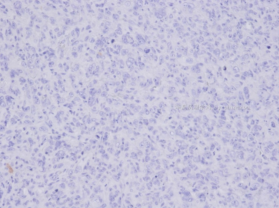
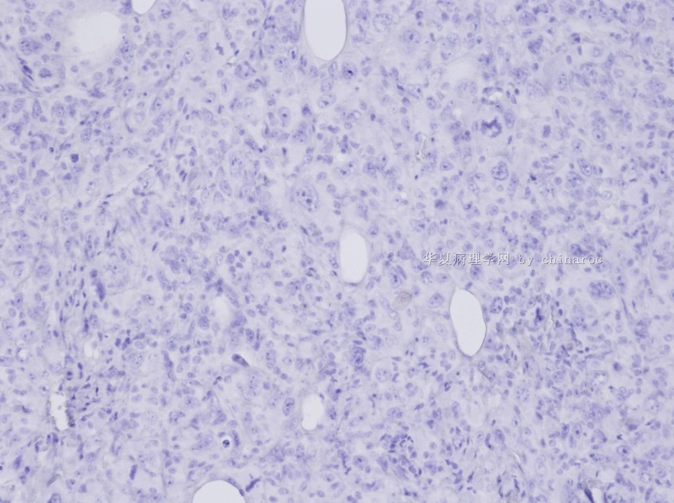
2)形态学符合多形性恶性肿瘤。
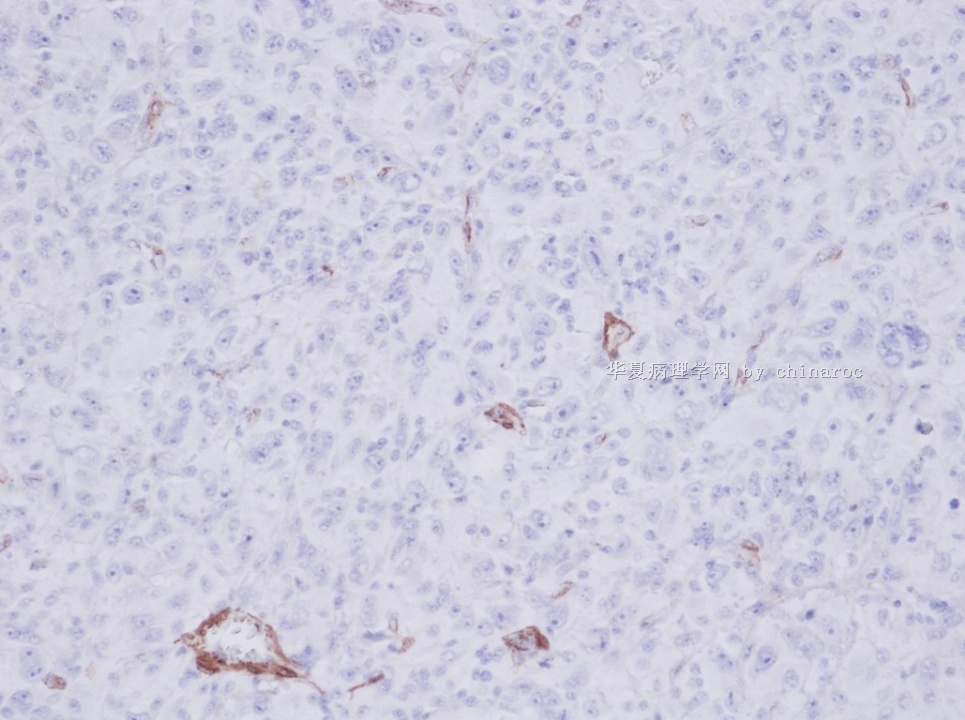
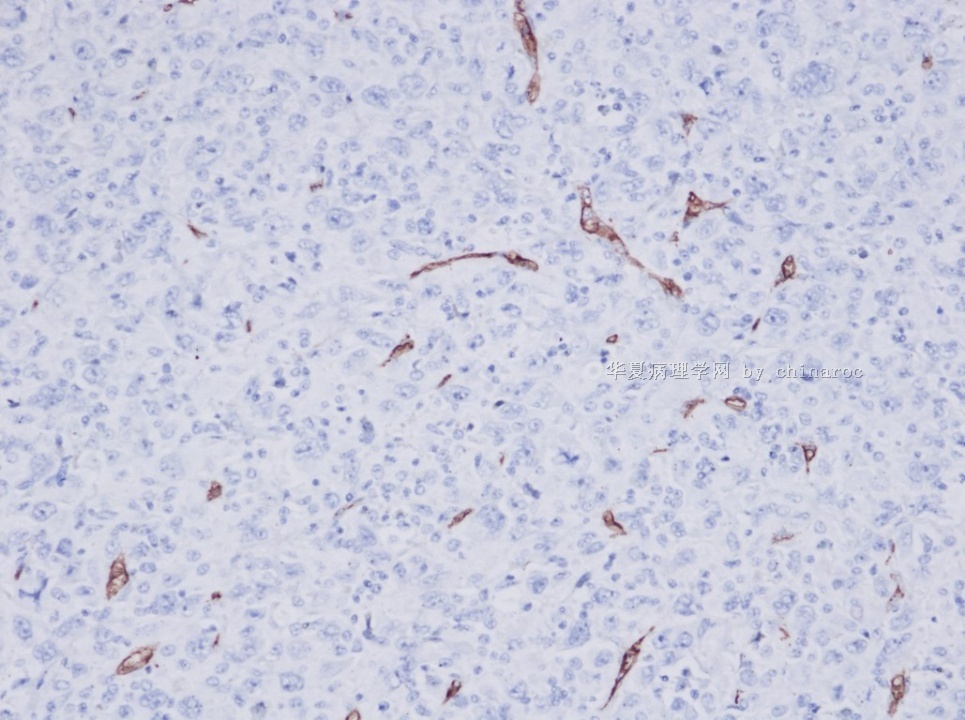
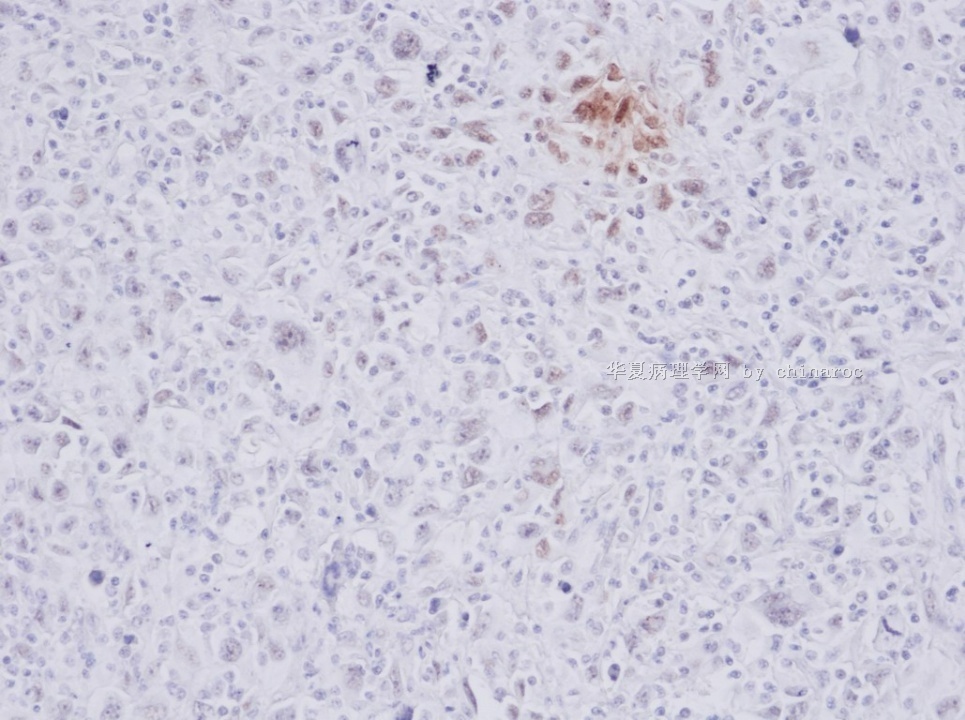
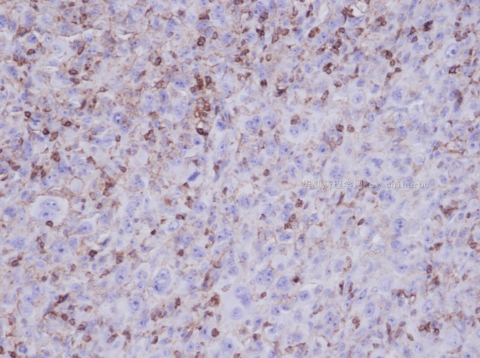
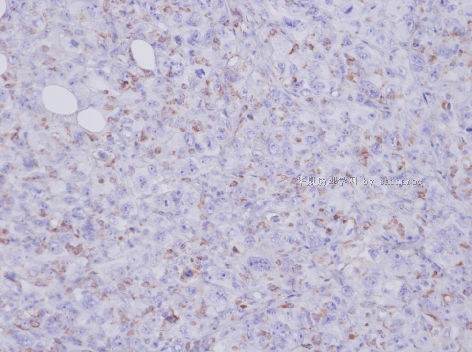
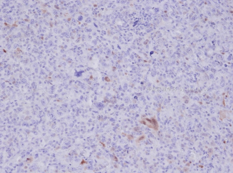
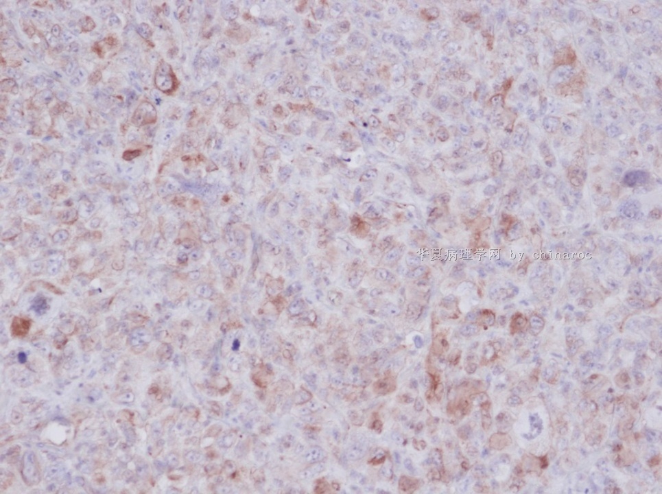
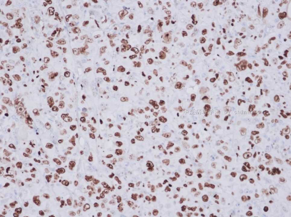
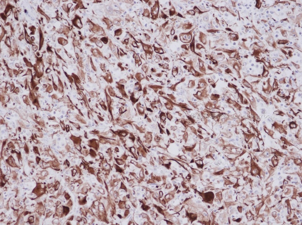
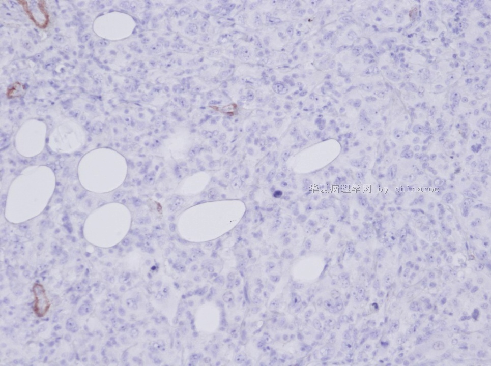
3)IHC标记:肿瘤细胞Cam5.2+Vimentin+GPC-3+、SMA-Desmin-CD34-间皮细胞-; 间质细胞LCA+CD68+。
鉴别诊断需考虑:
1)恶性间皮瘤,增加标记:CK5/6、Calretinin、WT-1、D2-40、P16;
2)GIST, 但是Cam5.2 +难解释。可增加标记除外:Dog-1、CD117、CD34、Nestin。
3)经典性HL, 淋巴细胞消减型。Cam-5.2+难解释。增加标记除外:CD15、CD30。
4)多形性未分化肉瘤/MFH。须除外其他类型后考虑。
5)转移性肉瘤样肝细胞癌(差分化肝细胞癌)。根据:Cam5.2+、GPC-3+。要了解血浆AFP、是否存在肝硬化、肝脏影像学。
鉴别诊断中最好增加标记:
HepPar1、 CEA-多抗、CK8/18、AFP、HMB-45、EMA、CEA、CDX-2。
谢谢!

- xljin8
-
本帖最后由 于 2010-12-09 06:46:00 编辑
| 以下是引用xljin8在2010-12-9 6:42:00的发言:
Shirakawa H, Suzuki H, Shimomura M, Kojima M, Gotohda N, Takahashi S, Nakagohri T, Konishi M, Kobayashi N, Kinoshita T, Nakatsura T. Glypican-3 expression is correlated with poor prognosis in hepatocellular carcinoma. Cancer Sci. 2009;100(8):1403-7.
The relationship between overexpression of glypican (GPC)-3 that is specific for hepatocellular carcinoma (HCC) and the prognosis has not yet been clarified. We attempted to determine the expression profile of GPC3 in association with the clinicopathological factors by immunohistochemical analysis in HCC patients and investigated the potential prognostic value of GPC3 by comparing the survival rate between the GPC3-positive and GPC3 negative HCC patients. Primary HCC tissue samples (n = 107)obtained from patients who had undergone hepatectomy between 2000 and 2001 were analyzed. GPC3 expression was less frequently observed in well-differentiated HCC than in moderately and poorly differentiated HCC, the difference in the frequency being statistically significant. GPC3-positive HCC patients had a significantly lower 5-year survival rate than the GPC3-negative HCC patients (54.5 vs 87.7%, P = 0.031). Among 80 of the 107 (74.6%) patients with initial treatment who underwent hepatectomy, none of GPC3-negative HCC patients (n = 16, 20.0%) died during the follow-up period. No deaths were noted in the GPC3-negative HCC patients among the 71 (88.7%) patients with moderately and poorly differentiated HCC. Multivariate analysis identified GPC3 expression (P = 0.034) as an independent prognostic factor for the overall survival. We showed that GPC3 expression is correlated with a poor prognosis in HCC patients. |
- xljin8
-
This is a high grade and undifferentiated metastatic carcinoma of non-small cell type and of unclear primary anatomic/histologic origin. I do not think this is metastatic hepatocellular carcinoma, alveolar soft part sarcoma, melanoma, or classic Hodgkin lymphoma. Dendritic cell sarcoma and malignant mesothelioma are unlikely, but deserve to be ruled out by immunohistochemistry. It is very difficult to guess the primary origin, since this type of undifferentiated malignancy can be seen rarely in many organs, including kidney, pancreas, thyroid, testicle, liver and lung. I appreciate your sharing this challenging case, and look forward to any pertinent follow-up.
聞道有先後,術業有專攻
-
huisheng97 离线
- 帖子:263
- 粉蓝豆:22
- 经验:285
- 注册时间:2009-02-13
- 加关注 | 发消息
| 以下是引用mjma在2010-12-9 14:13:00的发言: This is a high grade and undifferentiated metastatic carcinoma of non-small cell type and of unclear primary anatomic/histologic origin. I do not think this is metastatic hepatocellular carcinoma, alveolar soft part sarcoma, melanoma, or classic Hodgkin lymphoma. Dendritic cell sarcoma and malignant mesothelioma are unlikely, but deserve to be ruled out by immunohistochemistry. It is very difficult to guess the primary origin, since this type of undifferentiated malignancy can be seen rarely in many organs, including kidney, pancreas, thyroid, testicle, liver and lung. I appreciate your sharing this challenging case, and look forward to any pertinent follow-up. |
-
Epithelioid variant of pleomorphic liposarcoma: a comparative immunohistochemical and ultrastructural analysis of six cases with emphasis on overlapping features with epithelial malignancies.
Department of Pathology, Memorial Sloan-Kettering Cancer Center, New York, New York, USA.
Ultrastruct Pathol. 2002 Sep-Oct;26(5):299-308.
Abstract
Pleomorphic liposarcoma (PL) is the least common subtype of liposarcoma, displaying a lipoblastic, malignant fibrous histiocytoma (MFH)-like and, less frequently, an epithelioid growth pattern. The epithelioid morphology in PL is still underrecognized and may closely simulate other epithelial neoplasms, mainly adrenal cortical carcinoma (ACC). No electron microscopic (EM) studies of the epithelioid variant of PL have been previously described, nor have there been studies of its immunoreactivity with A103 or alpha-inhibin. The purpose of this study is to analyze the histological, immunohistochemical, and EM features of epithelioid PL in an attempt to better explore the distinction from their epithelial mimickers, such as ACC. A panel of 5 antibodies was studied, including A103, alpha-inhibin, smooth muscle actin (SMA), AE1/AE3, and Cam 5.2. Out of 22 cases of PLs, 6 cases characterized by the presence of both epithelioid phenotype and pleomorphic lipoblasts were identified from the EM archives. There were 4 females and 2 males, with a mean age of 58 (range, 39-78). Two lesions arose in the thigh and 1 each in the abdominal wall, chest wall, anterior mediastinum, and retroperitoneum, with tumor size ranging from 7 to 17 cm (mean, 13 cm). Histologically, 2 PLs were pure epithelioid, whereas the other 4 had a mixed epithelioid and MFH-like appearance. Immunohistochemically, A103 (4/6), SMA (4/6), and AE1/AE3 (1/6) revealed a various degree of positive reactions. No immunolabeling for alpha-inhibin or Cam5.2 was detected in any case. By EM, the epithelioid areas revealed round or polyhedral cells with lipid droplets of various sizes and number, intimately apposed cell surfaces, occasional junction-like structures (4/6), and micropinocytotic vesicles (4/6). Interestingly, the ribosome-lamellar complexes, once thought to be characteristic of hairy cell leukemia but rarely seen in solid tumors, were noted in one pure epithelioid PL. When compared to the MFH-like area, rough endoplasmic reticula (RER) were less well developed, but mitochondria were more prominent in the epithelioid components. Neither mitochondria with tubulovesicular cristae nor prominent smooth endoplasmic reticula indicative of ACC were seen. Well-formed external lamina was not present. Other features to support a higher level of epithelial differentiation, such as lumen formation, microvilli, and tonofilaments, were not found. In conclusion, focal A103 reactivity in epithelioid undifferentiated tumors should be interpreted with caution before rendering the diagnosis of a primary or metastatic ACC, especially when examining biopsy specimens. The possibility of an epithelioid variant of PL must be excluded; alpha-inhibin can serve as a useful adjunct in this regard. In addition to variable intracytoplasmic fat droplets, the distinctive ultrastructural features of epithelioid variant of PL include numerous mitochondria, pinocytotic vesicles, junction-like structures, and, rarely, ribosome-lamellar complex. Despite some overlapping features, electron microscopy remains a useful tool to distinguish between epithelioid PL and ACC.
- 用心做事、真情做人!


