| 图片: | |
|---|---|
| 名称: | |
| 描述: | |
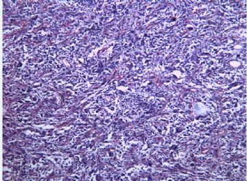
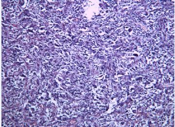
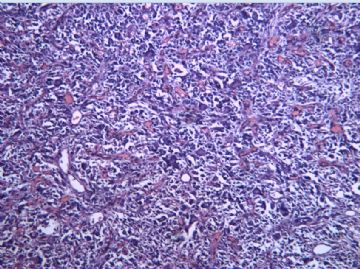
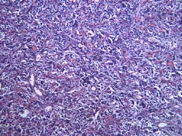
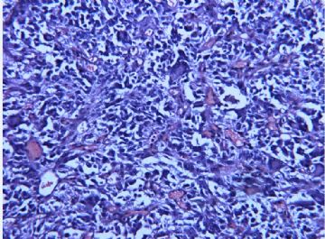
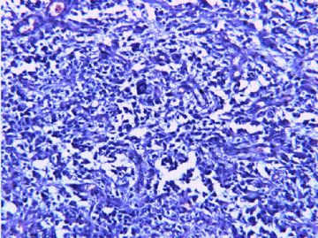
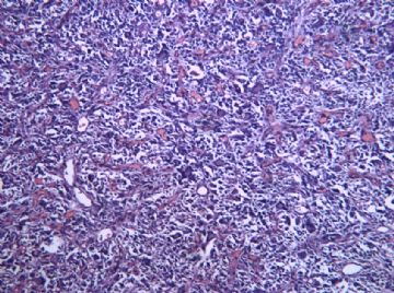
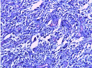
- 脑室占位--多形性黄色星型细胞瘤?
-
The photos do suggest a case of pleomorphic xanthoastrocytoma. Please search for eosinophilic granular bodies and perivascular inflammation on sections carefully. If both are present the diagnosis is WHO grade II PXA. In addition, mitotic count is very important because very rare cases of PXA behave more aggressively and their only atypical histologic features are increased mitotic activity and focal tumor necrosis. Of course, one important differential diagnosis is giant cell variant of glioblastoma.

聞道有先後,術業有專攻

