| 图片: | |
|---|---|
| 名称: | |
| 描述: | |
- 右顶叶占位
-
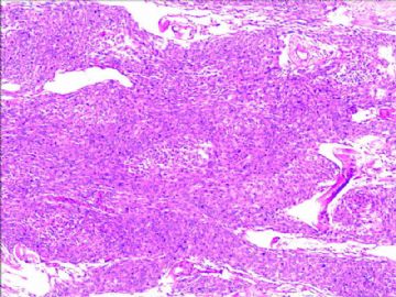
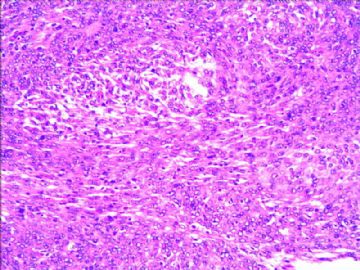
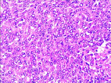
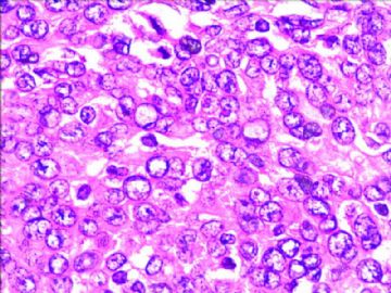
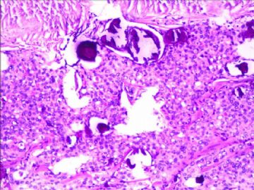
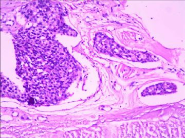
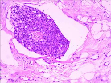
This is a meningioma of meningothelial type. Grading requires the examiner to carefully count mitoses and to find the following atypical features - necrosis, prominent nucleoli, small cells with high nucleocytoplasmic ratios, hypercellularity and sheet-like growth. If mitotic count is greater or equal to 4 per 10 hpf, this would qualify as WHO grade II atypical meningioma. If three or more of the five atypical features are present, this would also qualify as WHO grade II atypical meningioma. One should also try to see if there is any adherent brain parenchyma and whether the tumor has invaded into ti. Seeing tumor cell nests in the veins of dura is not a feature of atypical or malignant meningioma.

聞道有先後,術業有專攻
| 以下是引用mjma在2010-10-25 21:05:00的发言: This is a meningioma of meningothelial type. Grading requires the examiner to carefully count mitoses and to find the following atypical features - necrosis, prominent nucleoli, small cells with high nucleocytoplasmic ratios, hypercellularity and sheet-like growth. If mitotic count is greater or equal to 4 per 10 hpf, this would qualify as WHO grade II atypical meningioma. If three or more of the five atypical features are present, this would also qualify as WHO grade II atypical meningioma. One should also try to see if there is any adherent brain parenchyma and whether the tumor has invaded into ti. Seeing tumor cell nests in the veins of dura is not a feature of atypical or malignant meningioma. 这是一例上皮细胞型的脑膜瘤。它的分级需要诊断医生仔细计数核分裂相及以下非典型特征——坏死,显著的核仁,具有高核浆比的小细胞,细胞生长活跃,片状生长方式。如果和分裂相计数大于或等于4 /10 hpf就可以诊断为WHOⅡ级的非典型脑膜瘤。如果以上所述的5种非典型特征中存在3个或更多,也可以诊断为WHOⅡ级的非典型脑膜瘤。我们也应该努力寻找肿瘤是否粘附于脑组织,或已经浸润脑组织。脑膜静脉血管内可见肿瘤细胞巢不是非典型脑膜瘤或恶性脑膜瘤的特征。 |

