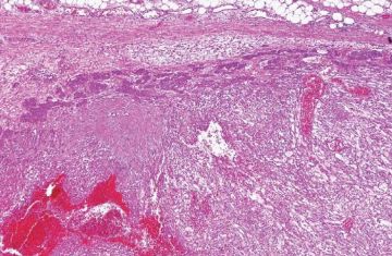
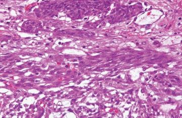
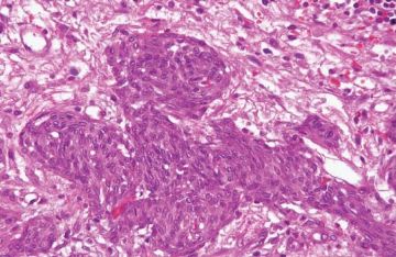
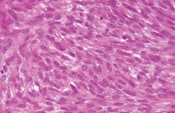
下列是两篇关于“胃母细胞瘤”的参考文献。这个诊断名词只是去年由Miettinen等提出,今年又报道1例,全球共报道4例。
组织学特点是双向分化。
IHC表型主要是上皮细胞CK(AE1/AE3+、CK18+和CK7+;而CK5/6和CK20是阴性的);梭形间叶细胞表达CD10+。
1. J Clin Pathol. 2010 Mar;63(3):270-4.
Novel epitheliomesenchymal biphasic stomach tumour (gastroblastoma) in a 9-year-old: morphological, ultrastructural and immunohistochemical findings.
Shin DH, Lee JH, Kang HJ, Choi KU, Kim JY, Park do Y, Lee CH, Sol MY, Park JH, Kim HY, Montgomery E.
Department of Pathology, Pusan National University School of Medicine, Yang-san, Korea.
Abstract
Gastroblastoma is a rare gastric epitheliomesenchymal biphasic tumour composed of spindle and epithelial cells, reported by Miettinen et al in a series of three cases in 2009. All those cases arose in stomachs of young adults. Neither the epithelial nor the mesenchymal component displayed sufficient atypia to diagnose a carcinosarcoma or other malignancy. On immunohistochemistry, the epithelial component expressed cytokeratin, and the mesenchymal component was positive for vimentin and CD10. Miettinen et al designated these neoplasms as gastroblastomas based on their similarities with other childhood blastomas such as pleuropulmonary blastoma and nephroblastoma. This report describes a probable fourth case of this unique type of neoplasm. The present case arose in the gastric antrum of a 9-year-old boy. While similarities were evident with the other cases, there were some differences. The epithelial component was more predominant and showed more mature morphology. Immunohistochemically, the epithelial component showed immunolabelling for c-KIT and CD56. The mesenchymal component was only focally positive for CD10. Ultrastructually, desmosomes and microvilli were found supporting a truly epithelial lesion.
2. Am J Surg Pathol. 2009 Sep;33(9):1370-7.
A distinctive novel epitheliomesenchymal biphasic tumor of the stomach in young adults ("gastroblastoma"): a series of 3 cases.
Miettinen M, Dow N, Lasota J, Sobin LH.
Department of Soft Tissue Pathology, Armed Forces Institute of Pathology, 6825 16th Street, N.W., Building 54, Room G090, Washington, DC 20306-6000, USA. miettinen@afip.osd.mil
Abstract
This report describes 3 cases of a distinctive, hitherto unreported gastric epitheliomesenchymal biphasic tumor that differs from other biphasic tumors of the stomach and elsewhere: carcinosarcoma, biphasic synovial sarcoma, teratoma, and mixed tumor. The tumors occurred in young adults, 2 males and 1 female, of ages 19, 27, and 30 years. Two tumors were located in the greater curvature in the gastric body and one in the antrum. The tumors measured 5, 6, and 15 cm in maximum diameter, and their mitotic rates were 0, 4, and 30 mitoses per 50HPF. There were 2 components: uniform oval or spindled cells in diffuse sheets, and clusters or cords of epithelial cells occasionally forming glandular structures with small lumens. The epithelial elements were positive for keratin cocktail AE1/AE3, keratin 18, and partly for keratin 7, but were negative for keratins 5/6, 20 and epithelial membrane antigen. The spindle cells were positive for vimentin and CD10. All components were negative for CD34, CD99, estrogen receptor, KIT, smooth muscle actin, desmin S100 protein, p63, calretinin, chromogranin, synaptophysin, CDX2, and thyroid transcription factor 1. In situ hybridization for SS18 rearrangement was negative in all cases separating this tumor from synovial sarcoma. All 3 patients were alive after follow-up of 3.5, 5, and 14 years. Because these tumors have some resemblance to blastomas of other organs, we propose the term "gastroblastoma" for this distinctive, at least low-grade malignant epitheliomesenchymal tumor of the stomach.
下列是两篇关于“胃母细胞瘤”的参考文献。这个诊断名词只是去年由
Miettinen等提出,今年又报道1例,全球共报道4例。
组织学特点是双向分化。
IHC表型主要是上皮细胞CK(AE1/AE3+、CK18+和CK7+;而CK5/6和CK20是阴性的);梭形间叶细胞表达CD10+。
1. J Clin Pathol. 2010 Mar;63(3):270-4.
Novel epitheliomesenchymal biphasic stomach tumour (gastroblastoma) in a 9-year-old: morphological, ultrastructural and immunohistochemical findings.
Shin DH, Lee JH, Kang HJ, Choi KU, Kim JY, Park do Y, Lee CH, Sol MY, Park JH, Kim HY, Montgomery E.
Department of Pathology, Pusan National University School of Medicine, Yang-san, Korea.
Abstract
Gastroblastoma is a rare gastric epitheliomesenchymal biphasic tumour composed of spindle and epithelial cells, reported by Miettinen et al in a series of three cases in 2009. All those cases arose in stomachs of young adults. Neither the epithelial nor the mesenchymal component displayed sufficient atypia to diagnose a carcinosarcoma or other malignancy. On immunohistochemistry, the epithelial component expressed cytokeratin, and the mesenchymal component was positive for vimentin and CD10. Miettinen et al designated these neoplasms as gastroblastomas based on their similarities with other childhood blastomas such as pleuropulmonary blastoma and nephroblastoma. This report describes a probable fourth case of this unique type of neoplasm. The present case arose in the gastric antrum of a 9-year-old boy. While similarities were evident with the other cases, there were some differences. The epithelial component was more predominant and showed more mature morphology. Immunohistochemically, the epithelial component showed immunolabelling for c-KIT and CD56. The mesenchymal component was only focally positive for CD10. Ultrastructually, desmosomes and microvilli were found supporting a truly epithelial lesion.
2. Am J Surg Pathol. 2009 Sep;33(9):1370-7.
A distinctive novel epitheliomesenchymal biphasic tumor of the stomach in young adults ("gastroblastoma"): a series of 3 cases.
Miettinen M, Dow N, Lasota J, Sobin LH.
Department of Soft Tissue Pathology, Armed Forces Institute of Pathology, 6825 16th Street, N.W., Building 54, Room G090, Washington, DC 20306-6000, USA. miettinen@afip.osd.mil
Abstract
This report describes 3 cases of a distinctive, hitherto unreported gastric epitheliomesenchymal biphasic tumor that differs from other biphasic tumors of the stomach and elsewhere: carcinosarcoma, biphasic synovial sarcoma, teratoma, and mixed tumor. The tumors occurred in young adults, 2 males and 1 female, of ages 19, 27, and 30 years. Two tumors were located in the greater curvature in the gastric body and one in the antrum. The tumors measured 5, 6, and 15 cm in maximum diameter, and their mitotic rates were 0, 4, and 30 mitoses per 50HPF. There were 2 components: uniform oval or spindled cells in diffuse sheets, and clusters or cords of epithelial cells occasionally forming glandular structures with small lumens. The epithelial elements were positive for keratin cocktail AE1/AE3, keratin 18, and partly for keratin 7, but were negative for keratins 5/6, 20 and epithelial membrane antigen. The spindle cells were positive for vimentin and CD10. All components were negative for CD34, CD99, estrogen receptor, KIT, smooth muscle actin, desmin S100 protein, p63, calretinin, chromogranin, synaptophysin, CDX2, and thyroid transcription factor 1. In situ hybridization for SS18 rearrangement was negative in all cases separating this tumor from synovial sarcoma. All 3 patients were alive after follow-up of 3.5, 5, and 14 years. Because these tumors have some resemblance to blastomas of other organs, we propose the term "gastroblastoma" for this distinctive, at least low-grade malignant epitheliomesenchymal tumor of the stomach.


