共2页/26条首页上一页12下一页尾页
回复:26 阅读:4350
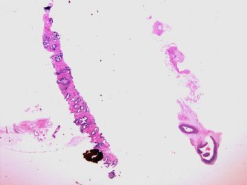
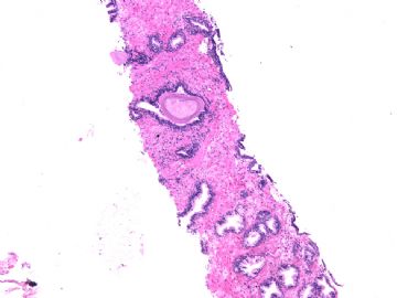
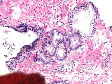
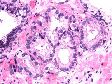
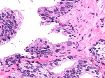
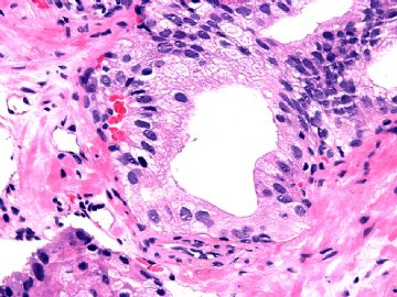
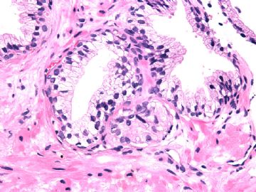
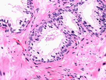
男/52岁 前列腺穿刺 诊断?

xljin8 离线

帖子:2275
粉蓝豆:1536
经验:2417
注册时间:2009-03-04
加关注  |  发消息
楼主 发表于 2010-10-06 18:57|举报|关注(1)
浏览排序[ 顺序 逆序 楼主 支持 精彩 ]  快捷回复
姓    名: ××× 性别:   年龄:  
标本名称:  
简要病史: 前列腺肥大,发现PSA升高2周。体检及B超前列腺无肿块。
肉眼检查:  
  • 男/52岁 前列腺穿刺 诊断?图1
    图1
  • 男/52岁 前列腺穿刺 诊断?图2
    图2
  • 男/52岁 前列腺穿刺 诊断?图3
    图3
  • 男/52岁 前列腺穿刺 诊断?图4
    图4
  • 男/52岁 前列腺穿刺 诊断?图5
    图5
  • 男/52岁 前列腺穿刺 诊断?图6
    图6
  • 男/52岁 前列腺穿刺 诊断?图7
    图7
  • 男/52岁 前列腺穿刺 诊断?图8
    图8
标签:
本帖最后由 于 2010-10-08 06:20:00 编辑
0
signature
xljin8
添加参考诊断
×参考诊断
  

xiaoming 离线

帖子:584
粉蓝豆:6
经验:688
注册时间:2008-07-18
加关注  |  发消息
1 楼    发表于2010-10-06 19:09:00举报|引用
返回顶部 | 快捷回复
 高级别PIN
0
回复

海上明月 离线

帖子:9476
粉蓝豆:1172
经验:10007
注册时间:2009-08-29
加关注  |  发消息
2 楼    发表于2010-10-06 19:16:00举报|引用
返回顶部 | 快捷回复
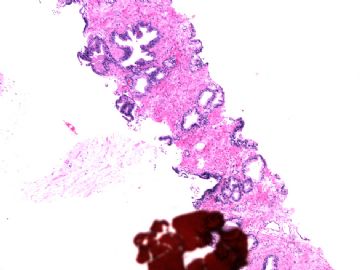
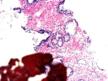
 关注标记处小灶非典型性腺泡。患者PSA升高。如果有足够的穿刺活检条数,看看他处是否还有这样的非典型性腺泡。
0
回复
signature
王军臣

shandongzhang 离线

帖子:804
粉蓝豆:506
经验:1510
注册时间:2006-09-24
加关注  |  发消息
3 楼    发表于2010-10-07 20:31:00举报|引用
返回顶部 | 快捷回复
 图3-4八个小灶非典型腺泡应该是癌,惜没有看到低倍相应所在部位,可能是另一条上的?
0
回复

XLJin8 离线

帖子:2275
粉蓝豆:1536
经验:2417
注册时间:2009-03-04
加关注  |  发消息
4 楼    发表于2010-10-07 21:20:00举报|引用
返回顶部 | 快捷回复
本帖最后由 于 2010-10-07 21:28:00 编辑


可疑病灶在图1上。增加图1点墨水处2张中间放大倍数照片。

  • 图1
  • 图2
0
回复
signature
xljin8

天山望月 离线

帖子:4902
粉蓝豆:83
经验:5092
注册时间:2007-02-03
加关注  |  发消息
5 楼    发表于2010-10-07 21:25:00举报|引用
返回顶部 | 快捷回复
 单层细胞,核仁明显,先看看IHC标记,很小范围,觉得还是非典型增生,建议再多穿刺。
0
回复
signature
广州金域病理

XLJin8 离线

帖子:2275
粉蓝豆:1536
经验:2417
注册时间:2009-03-04
加关注  |  发消息
6 楼    发表于2010-10-07 21:32:00举报|引用
返回顶部 | 快捷回复


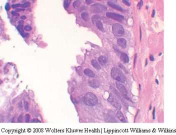
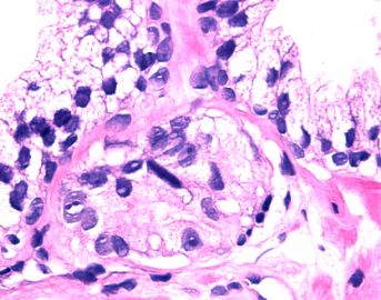
图5-8是可疑小腺体周围的腺泡,有高级别PIN改变,特别是图7。但是,基底细胞未完全消失。

0
回复
signature
xljin8

XLJin8 离线

帖子:2275
粉蓝豆:1536
经验:2417
注册时间:2009-03-04
加关注  |  发消息
7 楼    发表于2010-10-08 06:06:00举报|引用
返回顶部 | 快捷回复
本帖最后由 于 2010-10-08 06:13:00 编辑


请比较Tufting high-grade PIN

  • 图1
  • 图2
0
回复
signature
xljin8

XLJin8 离线

帖子:2275
粉蓝豆:1536
经验:2417
注册时间:2009-03-04
加关注  |  发消息
8 楼    发表于2010-10-08 06:37:00举报|引用
返回顶部 | 快捷回复
本帖最后由 于 2010-10-08 06:39:00 编辑


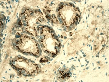
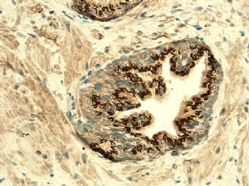
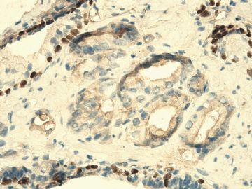
IHC P504S 图1可疑腺体、图2周围腺泡。

  • 图1
  • 图2
0
回复
signature
xljin8

XLJin8 离线

帖子:2275
粉蓝豆:1536
经验:2417
注册时间:2009-03-04
加关注  |  发消息
9 楼    发表于2010-10-08 08:14:00举报|引用
返回顶部 | 快捷回复
本帖最后由 于 2010-10-08 08:15:00 编辑

Epstein JI & Netto GJ.  

Biopsy interpretation of the prostate, 4th edtion,2008,p74.

表6-2  前列腺癌与PIN的鉴别诊断。

支持诊断癌 (Favoring Cancer)

反对诊断癌 (Against Cancer)

结构(Architectural)

浸润性构型

小腺体

腺体拥挤

细胞核(Nuclear)

核仁明显

核增大

核分裂

凋亡小体

细胞质(Cytoplasmic)

无定形细胞质

腔缘胞质整齐

管腔内容物

蓝色黏液性分泌物

粉红色无定形分泌物

结晶体

结构(Architectural)

小叶状结构

大腺体

分支腺体

细胞核(Nuclear)

核仁明显但是临近有PIN

核改变与邻近良性腺体相似

核不典型伴有炎症反应


细胞质(Cytoplasmic)

淡然-透明胞质

腔缘波纹状或乳头状突起

管腔内容物


淀粉样小体

0
回复
signature
xljin8

天山望月 离线

帖子:4902
粉蓝豆:83
经验:5092
注册时间:2007-02-03
加关注  |  发消息
10 楼    发表于2010-10-08 23:06:00举报|引用
返回顶部 | 快捷回复

 谢谢金主任提供对比诊断!收藏了。

此例免疫组化如何?重新阅片,单层细胞小腺体周围是PIN,可能考虑癌+PIN,不知最后结果如何?请继续讨论。

0
回复
signature
广州金域病理

海上明月 离线

帖子:9476
粉蓝豆:1172
经验:10007
注册时间:2009-08-29
加关注  |  发消息
11 楼    发表于2010-10-09 01:03:00举报|引用
返回顶部 | 快捷回复
本帖最后由 于 2010-10-09 19:30:00 编辑  标记区共有8个半非典型性小腺泡构成小病灶,旁边有PIN。标记P504s阳性,形态上见大核仁,无基底细胞,高度疑癌。如穿刺条数未达到6-12条,建议重取活检。
0
回复
signature
王军臣

wang4160 离线

帖子:2256
粉蓝豆:16
经验:2617
注册时间:2007-01-31
加关注  |  发消息
12 楼    发表于2010-10-09 08:17:00举报|引用
返回顶部 | 快捷回复
 学习一下!
0
回复

wy1992 在线

帖子:4858
粉蓝豆:1
经验:7320
注册时间:2007-06-30
加关注  |  发消息
13 楼    发表于2010-10-09 18:27:00举报|引用
返回顶部 | 快捷回复
 高级别上皮内瘤变
0
回复
signature

朱正龙

yihua0412 离线

帖子:262
粉蓝豆:1
经验:683
注册时间:2010-07-28
加关注  |  发消息
14 楼    发表于2010-10-10 10:28:00举报|引用
返回顶部 | 快捷回复

 结果,出来了吗?

请告诉我,好吗?

0
回复

hu8afu 离线

帖子:1177
粉蓝豆:35
经验:1230
注册时间:2009-02-11
加关注  |  发消息
15 楼    发表于2010-10-10 10:43:00举报|引用
返回顶部 | 快捷回复
 前列腺增生
0
回复

海上明月 离线

帖子:9476
粉蓝豆:1172
经验:10007
注册时间:2009-08-29
加关注  |  发消息
16 楼    发表于2010-10-10 13:41:00举报|引用
返回顶部 | 快捷回复
 
  海上明月 
  • 文章:4828
  • 积分:24255
  • 经验:4910
  • 注册:2009-8-29 13:59
  • 发表于 2010-9-2 17:0 {资料} {好友} {短信} {引用} {快回} {编辑} {删除 56 楼 
    以下是引用shandongzhang在2010-9-1 14:34:00的发言:

     Should Pathologists Diagnose ‘‘Adenocarcinoma’’When They Encounter a Single Small Atypical
    Focus in a Prostate Biopsy?
    In Response:
    Both Letters to the Editor referring to our paper ‘‘Variability in diagnostic opinion among pathologists for
    single small atypical foci in prostate biopsies’’ challenge its recommendation to obtain second opinion of a specialist pathologist before making a diagnosis of adenocarcinoma on a focus, comprising
    5 or less atypical acini.4 Dr Dalton1 argues that the clinical outcome of men with these lesions, irrespective
    of the diagnosis, is any how favorable.His view seems to be further corroborated by another paper recently published in this Journal5 on false negative prostate biopsies that is, biopsies where the diagnosis of prostate cancer was missed. The missed lesions were small foci of Gleason score 6 (3 +3) denocarcinomas in a single biopsy. Indeed,none of the men with a false-negative biopsy seemed to have a cancer advanced beyond the curable stage at the time of prostate cancer diagnosis 4 to 8 years later and the cancers did not differ from those found in men with a previous truly negative biopsy result.
    To our opinion, the argument raised by Dr Dalton1 might be valid at a population level, but may not apply
    for the individual man, in whom the small atypical focus could represent the tip of the iceberg, requiring
    additional clinical investigations. As literature has shown that intercollegial consultation leads to more definite diagnoses of benign or cancer in prostate biopsies,3 we maintain our recommendation on this matter.The reason for the disagreement of Drs Gilks and Huntsmans2 with our position is the lack of consensus among the experts themselves. On account of this lack of agreement on the diagnosis
    of such small foci, no individual expert can function as a ‘‘golden standard’’and therefore, they propose that pathologists should refrain from making a definite diagnosis of cancer when the number of atypical acini does not exceed a given number, for example, 10 acini. In contrast, we would argue that not all lesions with fewer than 6 atypical acini in our study suffered from a lack of consensus among experts, and in average the experts did better than the general pathologists of our study. The proportion
    of consensus in these cases might in fact be higher in a real-life situation as compared with this virtual microscopy environment, for reasons outlined in the discussion of our paper. Thus, we do not agree with
    a recommendation that would deny a pathologist’s right to render a definite diagnosis of adenocarcinoma when he would feel comfortable enough to do so. Obviously, we endorse the view
    that the scientific community should continue to look for additional tools to reduce the diagnostic uncertainty on these small atypical foci. In the mean time, we would encourage pathologists
    to seek advice from specialist pathologists to reduce the uncertainty of their diagnosis under these circumstances.

    Theodorus H. Van der Kwast, MD,
    PhD*
    Jonathan I. Epstein, MD, PhDw
    *Department of Pathology University
    Health Network Toronto, Canada
    wDepartment of Pathology
    The Johns Hopkins Medical Institutions
    Baltimore, MD

    Van der Kwast 和 Epstein 教授对读者来信的回复,试译如下:

    读者来信:病理医生在前列腺活检中遇到单个非典型小病灶时应诊断为“腺癌”吗?

    答复: 对我们所著《病理医生之间对前列腺活检中单个非典型小病灶诊断意见分歧》一文,两封读者来信对文中的建议提出质疑。我们的文中建议在对仅有5个或5个以下的非典型性腺泡小病灶诊断为前列腺癌之前,要征询专科病理专家的意见。Dalton1医生在信中辩称,这些病变不论是否诊断为癌,其患者的临床预后无论如何都是良好的。他的这种观点在本杂志最近发表的其它文章中好像进一步被认可,其文中论及前列腺活检诊断假阴性的问题,即前列腺癌活检漏诊的问题。漏诊的病变为单个活检组织中Gleason 评分为 6 (3 +3)的小灶前列腺癌。 事实上,假阴性活检病例在4—8年后诊断前列腺癌时,没有哪一例似是不可治愈的进展期前列腺癌,而且与先期活检确诊为真阴性的患者的发病情况没有什么不同之处。

     我们认为,Dalton1提出的论点在整体层面上可能是合理的,但具体应用到个体层面可能是不合适的。非典型性小病灶代表冰山一角,需要再做一些其它的临床检查工作。 正如文献所云,同行间会诊会使前列腺活检中良性或癌的诊断更加准确,为此,我们继续坚持我们文中的建议意见。Gilks 和 Huntsmans医生不同意我们的立场,其原因是其专家们自身之间没有达成共识。 由于对这种小病灶的诊断意见没有达成一致意见,无论哪一个专家的意见都不可以作为“金标准”。因此他们建议,当非典型性腺泡的数目不超过假定的数目(如给定10个腺泡)时,病理医生应避免确诊为癌。相反,我们的研究证明,即便是少于6个非典型性腺泡的病变,在我们的专家之间并不是对所有病变都缺乏共识意见,一般来说,专科病理专家要比参与我们研究的普通病理专家作出诊断的一致性更好些。与模拟显微镜诊断环境相比,在真实工作情景下对这些病变诊断一致性的比例要高得多。其原因在我们文章的讨论中已经作了概述。因此,我们不同意在足以诊断前列腺癌的时候而拒绝病理医生提出确诊意见权利的建议。 显然,我们赞同科学社团应继续寻找其他的评价方法,以减少对这些非典型性小病灶诊断的不确定性。同时,我们将鼓励病理医生向专科病理专家咨询,以减少其对这些非典型性小病灶诊断的不确定性。

    Theodorus H. Van der Kwast, MD,PhD*
    Jonathan I. Epstein, MD, PhDw
    *Department of Pathology University
    Health Network Toronto, Canada
    wDepartment of Pathology
    The Johns Hopkins Medical Institutions
    Baltimore, MD

    0
    回复
    signature
    王军臣

    海上明月 离线

    帖子:9476
    粉蓝豆:1172
    经验:10007
    注册时间:2009-08-29
    加关注  |  发消息
    17 楼    发表于2010-10-10 13:44:00举报|引用
    返回顶部 | 快捷回复
     请仔细阅读上楼的Epstein教授给读者的信。
    0
    回复
    signature
    王军臣

    shandongzhang 离线

    帖子:804
    粉蓝豆:506
    经验:1510
    注册时间:2006-09-24
    加关注  |  发消息
    18 楼    发表于2010-10-12 07:40:00举报|引用
    返回顶部 | 快捷回复
    以下是引用海上明月在2010-10-10 13:44:00的发言:

     请仔细阅读上楼的Epstein教授给读者的信。

    一字一句的阅读了N遍,仍在阅读中
    0
    回复

    XLJin8 离线

    帖子:2275
    粉蓝豆:1536
    经验:2417
    注册时间:2009-03-04
    加关注  |  发消息
    19 楼    发表于2010-10-12 17:58:00举报|引用
    返回顶部 | 快捷回复
    本帖最后由 于 2010-10-13 15:41:00 编辑

    对于活检组织中有少量异常腺体时要给予一个非常肯定的诊断有时是非常困难的, 这不仅在前列腺活检、同样包括乳腺、GI、呼吸道等器官的活检。

    解决疑难的方法:

    1)密切联系临床, 与临床医生沟通;

    2)详细分析对病理诊断有帮助的实验室数据和影像学资料等;

    3)仔细的镜下观察,与其他病理医生讨论;

    4)完善病理诊断必须的临床资料;

    5)合理选择IHC标记;

    6)如不能明确诊断,与病家沟通,建议随访、重复活检、专家会诊。


    病理医生要区分活检组织中的少量可疑病变可能存在的二种基本情况:

    1)患有是进展期癌,只不过活检组织中存在微量的癌(冰山一角);

    2)患者是局灶性癌前期病变(高级别PIN/上皮内瘤变)或微量早癌。


    病理医师要了解自己的诊断对临床处理产生的影响、了解对病人疾病预后与生活质量产生的影响。不要拒绝请专家会诊, “Second Lock” 有助于病人、有助于初诊医生的业务提高,何乐而不为? 病理诊断是艺术!有时退一步(不勉强诊断和过诊断)海阔天空。

    0
    回复
    signature
    xljin8

    XLJin8 离线

    帖子:2275
    粉蓝豆:1536
    经验:2417
    注册时间:2009-03-04
    加关注  |  发消息
    20 楼    发表于2010-10-13 15:57:00举报|引用
    返回顶部 | 快捷回复
    本帖最后由 于 2010-10-19 17:26:00 编辑


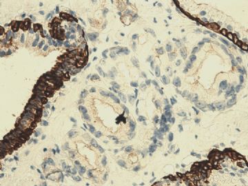
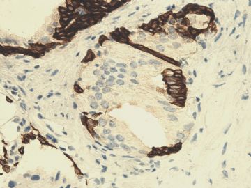
    IHC标记 34BE12 图1 可疑不病灶;图2 PIN;图3 p63。

    • 图1
    • 图2
    • 图3
    0
    回复
    signature
    xljin8
    回复:26 阅读:4350
    共2页/26条首页上一页12下一页尾页
    【免责声明】讨论内容仅作学术交流之用,不作为诊疗依据,由此而引起的法律问题作者及本站不承担任何责任。
    快速回复
    进入高级回复
    您最多可输入10000个汉字,按 "Ctrl" + "Enter" 直接发送
    搜索回复/乘电梯 ×
    按内容
    按会员
    乘电梯
    合作伙伴
    友情链接