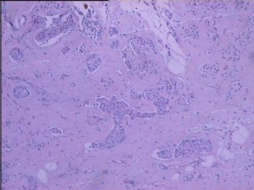
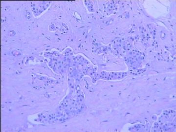
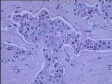
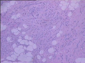
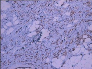
| 图片: | |
|---|---|
| 名称: | |
| 描述: | |
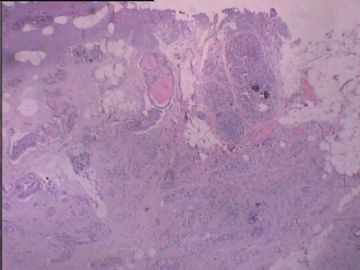
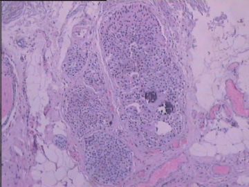
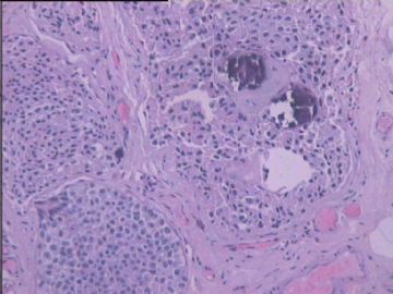
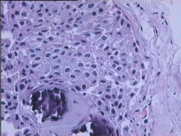
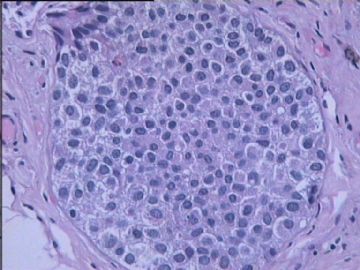
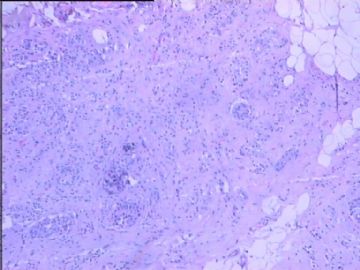
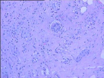
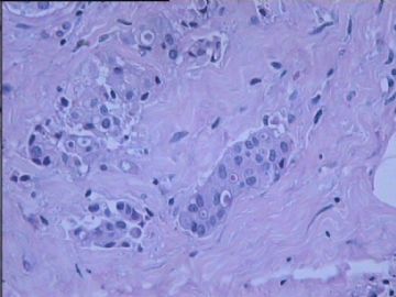
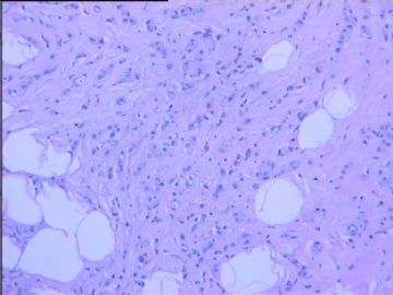
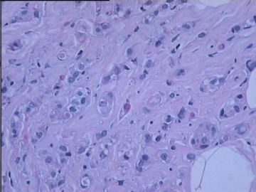
- B2914乳腺浸润性导管癌?
| 姓 名: | ××× | 性别: | 女 | 年龄: | 57 |
| 标本名称: | 左侧乳腺包块 | ||||
| 简要病史: | 发现左侧乳腺包块1年+。查体:左侧乳腺外上象限扪及一包块,直径约1.5cm,手术切除送检 | ||||
| 肉眼检查: | |||||
相关帖子
- • 乳腺肿物
- • 乳腺癌吗??
- • 左乳肿块,协助诊断
- • 左乳肿块
- • 乳腺肿物
- • 乳腺小管癌?
- • 左乳肿瘤--浸润性导管癌?
- • 看看这是那个类型的乳腺癌?
- • 乳腺肿物,请大家帮忙会诊是恶性的吗??
- • 急`1`1`1`1乳腺肿物,请大家帮忙会诊
| 以下是引用cqzhao在2010-10-13 4:15:00的发言:
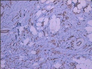
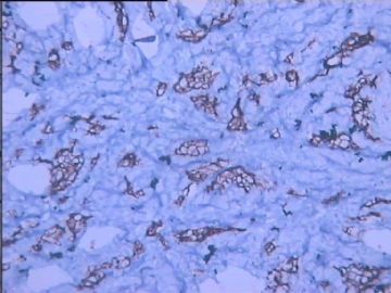
May be ductal ca. However I cannot evaluate if the stains of tumor cells are weaker than normal ductal epithelial cells based on photos. Remember that E-cadherin can be negative or reduced stain for E-cad |
-
wangdingding 离线
- 帖子:1474
- 粉蓝豆:98
- 经验:6042
- 注册时间:2006-10-19
- 加关注 | 发消息
It may be a ductal lesion. This is why we need to stain.
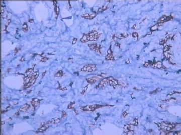
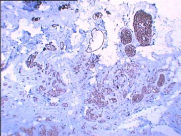
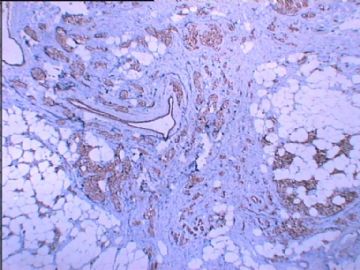
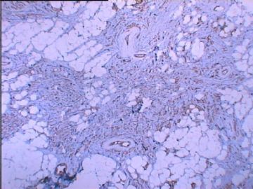
E-cad is membranous stain for ductal lesions. Your photos of stains are not clear. Can you show us the photos with high power and high quality? Do you have negative control? Can you do P120 stain?
thanks
| 以下是引用byq在2010-10-6 8:38:00的发言: 谢谢赵老师!谢谢强子翻译!对这个病例我们是有争论的,说实在话,我不是很肯定它一定就是癌,尤其是浸润性癌,因此我选择了CK5/6,SMA、P63,染CD34是因为我觉得第8-11副图中像肿瘤细胞在脉管内(我没有D2-40),不知道这样做是否合适?请赵老师指正。谢谢! |
Iy you are not sure, it is reasonable for the stains.
CD31 may be better than CD34. The specificity of CD34 is low. Should have D2-40 stains in your lab
综合cqzhao老师的回复:
做免疫组化要有一定的考虑,为什么选择上述指标(ER、PR、E-cadherin,CK5/6,P63,SMA,CD34)呢?
该例含有浸润性成分及原位成分,因此免疫组化CK5/6, sma, p63 并非必需,并且为什么染CD34呢?
当然,E-cad是为了区分导管癌及小叶癌, ER, PR, Her2 是为治疗
我的诊断:
浸润性小叶癌,组织学分级2级(小管形成-3,核级-2,核分裂-1,总6/9)
浸润性肿瘤大小:xx mm
小叶原位癌,经典型,核级-2,伴微钙化
小叶原位癌伴浸润性成分.
淋巴结浸润有/无
切缘阴性,浸润性癌距离切缘最近为xx mm
无肿瘤的乳腺组织见:xx, xx, xxxx
浸润性肿瘤部分 ER/PR/Her2 结果:

- 赚点散碎银子养家,乐呵呵的穿衣吃饭
My dx:
Invasive lobular ca, histological grade 2 (tubular formation-3, nuclear grade-2, mitotic activity-1; total score 6/9)
Invasive tumor measures xx mm
Lobular ca in situ, classic type, nuclear grade 2, with microcalcification
Lobular ca in situ mixed with invasive component.
No lymphovascular invasion present/or present.
Margins are negative for invasive ca; In vasive tumor is xx mm to the clostest xx margin
Non-neoplastic breast tissue showing xx, xx, xxxx
Invasive tumor ER/PR/Her2 results

