| 图片: | |
|---|---|
| 名称: | |
| 描述: | |
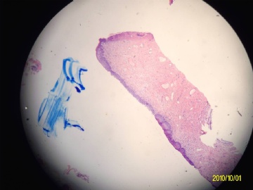
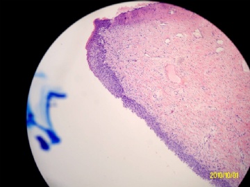
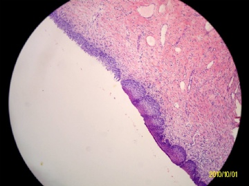
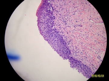
- 宫颈锥切组织
-
medman_2010 离线
- 帖子:402
- 粉蓝豆:1
- 经验:1245
- 注册时间:2009-05-13
- 加关注 | 发消息
非常遗憾,在免疫组化的片子已经看不到这片区域了,只能建议定期复查。
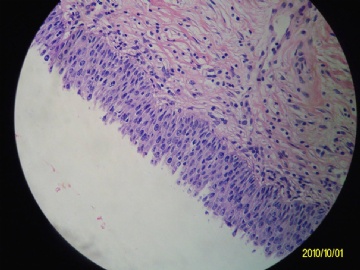
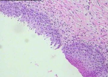
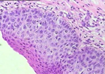
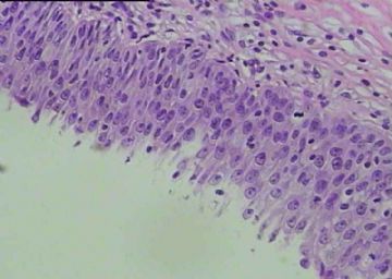
This is a very large tissue fragment. The materials should be more than enough to do 20 stains. Histotechnicians did not cut the slides well. Pathologists should give the patient or clinician a definite diagnosis for this case, but in fact we did not. This is a good example that 病理诊断是很严谨的,需多方合作,任一环节出问题,都可影响诊断。
| 以下是引用cqzhao在2010-10-3 20:18:00的发言:
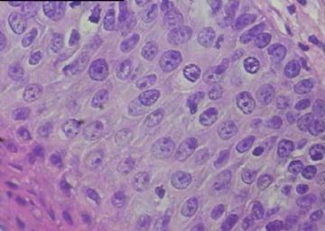
Cells look atypical ans show mitotic acitivity in the top 1/3 layer. It may be cin2-3. However cytologic features are not like classic high grade dysplasia, such as cell arrangement in order, nucleoli. This is why people above had different oppinions. P16 and Ki67 stains should be performed for this case. We are looking for your stains. thanks |
非常遗憾,在免疫组化的片子已经看不到这片区域了,只能建议定期复查。
非常感谢各位老师讨论。

- 简简单单,快快乐乐。
-
nfykdx2008 离线
- 帖子:734
- 粉蓝豆:28
- 经验:761
- 注册时间:2010-09-08
- 加关注 | 发消息
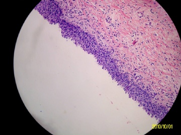
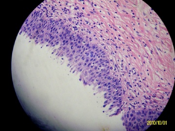
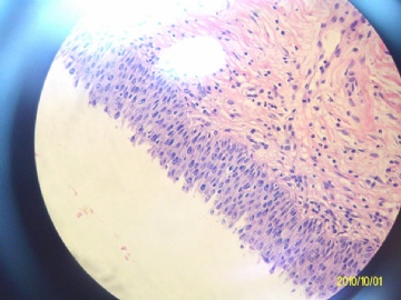
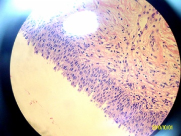
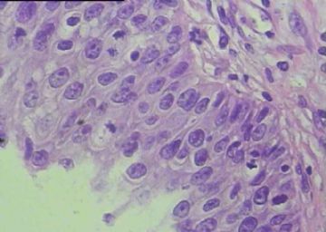
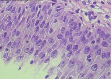
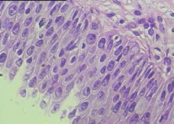
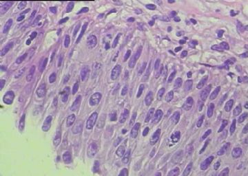
Cells look atypical ans show mitotic acitivity in the top 1/3 layer. It may be cin2-3. However cytologic features are not like classic high grade dysplasia, such as cell arrangement in order, nucleoli. This is why people above had different oppinions.
P16 and Ki67 stains should be performed for this case. We are looking for your stains. thanks

