
![[已确诊] 肺肿瘤_1 (lung tumor-1)- malignant melanoma 恶性黑色素瘤图1](/sites/default/uploads/old/2007-9/user_filesuserfile19358.jpg "[已确诊] 肺肿瘤_1 (lung tumor-1)- malignant melanoma 恶性黑色素瘤图1")
![[已确诊] 肺肿瘤_1 (lung tumor-1)- malignant melanoma 恶性黑色素瘤图2](/sites/default/uploads/old/2007-9/user_filesuserfile19359.jpg "[已确诊] 肺肿瘤_1 (lung tumor-1)- malignant melanoma 恶性黑色素瘤图2")
![[已确诊] 肺肿瘤_1 (lung tumor-1)- malignant melanoma 恶性黑色素瘤图3](/sites/default/uploads/old/2007-9/user_filesuserfile19360.jpg "[已确诊] 肺肿瘤_1 (lung tumor-1)- malignant melanoma 恶性黑色素瘤图3")
![[已确诊] 肺肿瘤_1 (lung tumor-1)- malignant melanoma 恶性黑色素瘤图4](/sites/default/uploads/old/2007-9/user_filesuserfile19361.jpg "[已确诊] 肺肿瘤_1 (lung tumor-1)- malignant melanoma 恶性黑色素瘤图4")
![[已确诊] 肺肿瘤_1 (lung tumor-1)- malignant melanoma 恶性黑色素瘤图5](/sites/default/uploads/old/2007-9/user_filesuserfile19362.jpg "[已确诊] 肺肿瘤_1 (lung tumor-1)- malignant melanoma 恶性黑色素瘤图5")
![[已确诊] 肺肿瘤_1 (lung tumor-1)- malignant melanoma 恶性黑色素瘤图6](/sites/default/uploads/old/2007-9/user_filesuserfile19363.jpg "[已确诊] 肺肿瘤_1 (lung tumor-1)- malignant melanoma 恶性黑色素瘤图6")
![[已确诊] 肺肿瘤_1 (lung tumor-1)- malignant melanoma 恶性黑色素瘤图7](/sites/default/uploads/old/2007-9/user_filesuserfile19366.jpg "[已确诊] 肺肿瘤_1 (lung tumor-1)- malignant melanoma 恶性黑色素瘤图7")


| 图片: | |
|---|---|
| 名称: | |
| 描述: | |
- [已确诊] 肺肿瘤_1 (lung tumor-1)- malignant melanoma 恶性黑色素瘤
![[已确诊] 肺肿瘤_1 (lung tumor-1)- malignant melanoma 恶性黑色素瘤图1](/sites/default/uploads/old/2007-9/small_user_filesuserfile19358.jpg "[已确诊] 肺肿瘤_1 (lung tumor-1)- malignant melanoma 恶性黑色素瘤图1") 图1
图1![[已确诊] 肺肿瘤_1 (lung tumor-1)- malignant melanoma 恶性黑色素瘤图2](/sites/default/uploads/old/2007-9/small_user_filesuserfile19359.jpg "[已确诊] 肺肿瘤_1 (lung tumor-1)- malignant melanoma 恶性黑色素瘤图2") 图2
图2![[已确诊] 肺肿瘤_1 (lung tumor-1)- malignant melanoma 恶性黑色素瘤图3](/sites/default/uploads/old/2007-9/small_user_filesuserfile19360.jpg "[已确诊] 肺肿瘤_1 (lung tumor-1)- malignant melanoma 恶性黑色素瘤图3") 图3
图3![[已确诊] 肺肿瘤_1 (lung tumor-1)- malignant melanoma 恶性黑色素瘤图4](/sites/default/uploads/old/2007-9/small_user_filesuserfile19361.jpg "[已确诊] 肺肿瘤_1 (lung tumor-1)- malignant melanoma 恶性黑色素瘤图4") 图4
图4![[已确诊] 肺肿瘤_1 (lung tumor-1)- malignant melanoma 恶性黑色素瘤图5](/sites/default/uploads/old/2007-9/small_user_filesuserfile19362.jpg "[已确诊] 肺肿瘤_1 (lung tumor-1)- malignant melanoma 恶性黑色素瘤图5") 图5
图5![[已确诊] 肺肿瘤_1 (lung tumor-1)- malignant melanoma 恶性黑色素瘤图6](/sites/default/uploads/old/2007-9/small_user_filesuserfile19363.jpg "[已确诊] 肺肿瘤_1 (lung tumor-1)- malignant melanoma 恶性黑色素瘤图6") 图6
图6![[已确诊] 肺肿瘤_1 (lung tumor-1)- malignant melanoma 恶性黑色素瘤图7](/sites/default/uploads/old/2007-9/small_user_filesuserfile19366.jpg "[已确诊] 肺肿瘤_1 (lung tumor-1)- malignant melanoma 恶性黑色素瘤图7") 图7
图7
| 姓 名: | ××× | 性别: | 60 | 年龄: | 男 male |
| 标本名称: | 肺叶切除术 lobectomy | ||||
| 简要病史: | 4厘米大肺肿瘤, 无已知恶性肿瘤病史 4 cm lung mass, no other known history of malignancy | ||||
| 肉眼检查: | 同上 | ||||
鉴别诊断, 诊断? 如果需要做核面议组化?
Your differential diagnosis and diagnosis? How to work it out if you need immunohistochemical stains?
标签:
-
本帖最后由 于 2007-10-06 09:30:00 编辑
×参考诊断
I agree 古城's observasion.
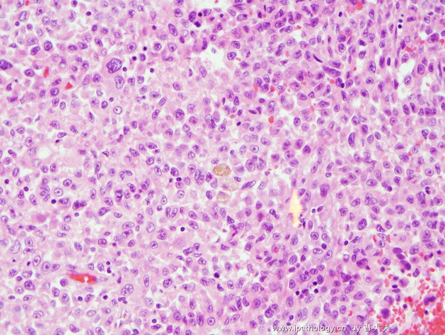
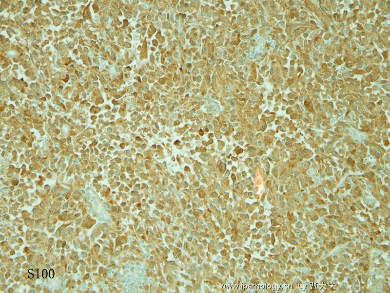
It is a quite circumscribed tumor formed by sheets of pleomorphic cells. In areas it has a papillary or rather pseudopapillary structure, and in areas it shows large solid sheets. the tumor cells are large epithelioid, with plenty eosinophilic cytoplasm, vesicular nuclei, and prominent nucleoli. many tumor cells show eccentric nuclei, some are indented due to abundent eosinophilic cytoplasm---rhabdoid morphology. multinucleated giant cells are present, and so as mitotic figures. there is small amount of inflammatory cells. no pigment is seen in thses slides.
consider patient's age, primary lung cancer has to be considered, a large cell carcinoma of lung would be my choice. secondary tumor such as metastatic melanoma should be in the differential diagnosis. other metastatic tumor such as carcinoma of other site or sarcoma, although less possible, also need to be excluded.
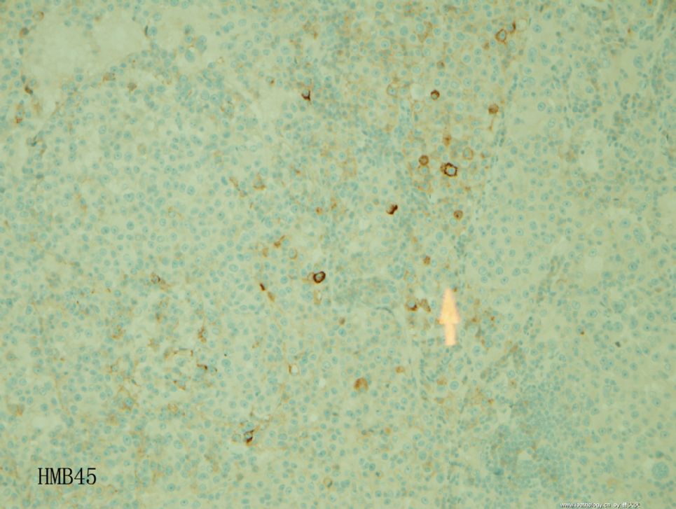
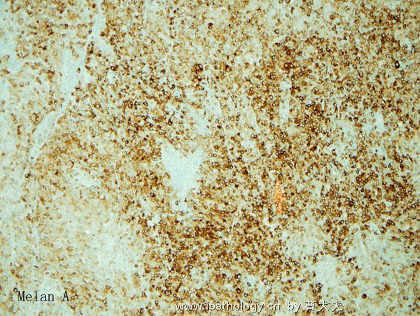
I would do: AE1/AE3, TTF1, vimentin, s-100, HMB45, Melanin A
-
本帖最后由 于 2007-10-06 09:33:00 编辑
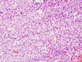 图1
图1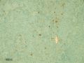 图2
图2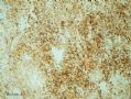 图3
图3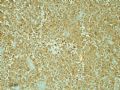 图4
图4
这个肿瘤是恶性黑色素瘤
这个病人其他地方没有恶性黑色素瘤病史,有2种可能:一是原发地已经退化但是肺有转移, 二是肺原发性恶性黑色素瘤(有过报道, 但是罕见, 这个病人的肺病变没有发现原位的黑色素瘤).
细胞当然有特点提示恶性黑色素瘤是在鉴别诊断中. 细胞相对松散, 有横纹肌肉瘤样的细胞, 有些地方看起来象有乳头样结构, 可能是中间的细胞松散所引起.
最后有一个地方有黑色素 (4 厘米大地肿瘤我们取了6个肿瘤片, 只有一个视野有黑色素). 开始我当然没有贴出来, 否则答案很快就明白了. 问题是当你看片时怎么找到这个视野.
The diagnosis for this case is malignant melanoma.
This patient does not have a history of malignant melanoma in other body sites. There are 2 possibilities for the lesion in the lung. First, the patient has a melanoma somewhere else but regressed and he developed a metastatic lesion in the lung. Second, primary lung melanoma has been reported but it is very rare. In this case we do not see any evidence of in situ melanoma.
The tumor cells have some characteristic features. They are somewhat discohesive and many of them have a rhabdoid appearance: eccentric nuclei with prominent nucleoli, abundant eosinophilic cytoplasm. In some areas there are some papille-like structures, probably caused by the discohesive cells between.
Melanin pigment is seen in only one area in this tumor. For a 4 cm mass we submitted 6 sections. This is the only area containing melanin pigment. I did not put this picture on at the beginning. Otherwise the answer will be so obvious. On the other hand, the key thing is how to find this field on the 6 slides.


