| 图片: | |
|---|---|
| 名称: | |
| 描述: | |
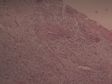
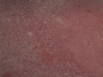
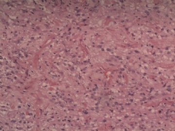
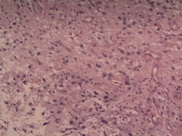
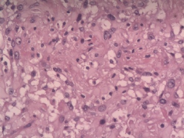
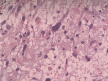
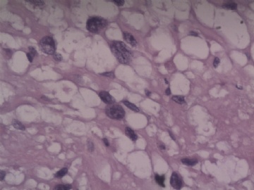
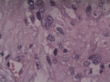
- 颞叶脑组织
-
These cells in brain parenchyma surrounding hemorrhage have atypical nuclei that makes me suspect an infiltrating astrocytoma. Is there any history of brain tumor before hemorrhage? Please see if more and better quality photos can be obtained and uploaded. GFAP, p53 and MIB-1 immunostains would help. Also do pay attention to small blood vessels in cortex and see if there is any features of cerebral amyloid angiopathy. Recurrent intracerebral hemorrhage in elderly should always raise this possibility.

聞道有先後,術業有專攻
| 以下是引用mjma在2010-9-29 22:27:00的发言: These cells in brain parenchyma surrounding hemorrhage have atypical nuclei that makes me suspect an infiltrating astrocytoma. Is there any history of brain tumor before hemorrhage? Please see if more and better quality photos can be obtained and uploaded. GFAP, p53 and MIB-1 immunostains would help. Also do pay attention to small blood vessels in cortex and see if there is any features of cerebral amyloid angiopathy. Recurrent intracerebral hemorrhage in elderly should always raise this possibility. |
Thank you for making a penetrating analysis
| 以下是引用mjma在2010-9-29 22:27:00的发言: These cells in brain parenchyma surrounding hemorrhage have atypical nuclei that makes me suspect an infiltrating astrocytoma. Is there any history of brain tumor before hemorrhage? Please see if more and better quality photos can be obtained and uploaded. GFAP, p53 and MIB-1 immunostains would help. Also do pay attention to small blood vessels in cortex and see if there is any features of cerebral amyloid angiopathy. Recurrent intracerebral hemorrhage in elderly should always raise this possibility. |
