| 图片: | |
|---|---|
| 名称: | |
| 描述: | |
- 看病理,学英语
-
本帖最后由 于 2010-09-18 12:14:00 编辑
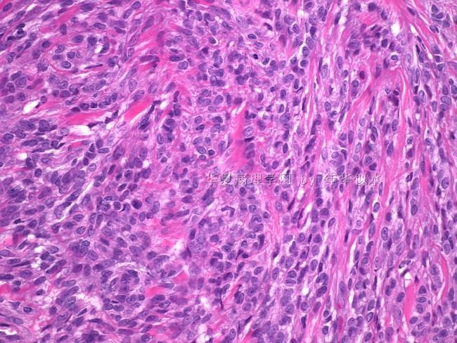
Epithelioid myofibroblastomas tend to be cellular and contain epithelioid cells arranged in small alveolar clusters or bundles. The main differential diagnosis is with invasive carcinoma and myoepithelioma.

名称:图1
描述:图1

- stay hungry,stay foolish.
Myofibroblastoma of breast
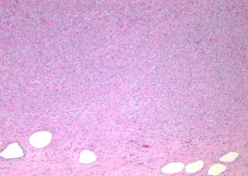
1,Well circumscribed
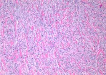
2, Uniform, bland spindle cells haphazardly arranged in fascicles with pushing borders, separated by broad bands of hyalinized collagen
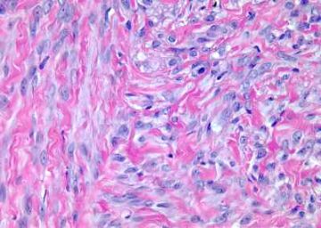
3,Spindle cells have abundant eosinophilic cytoplasm, round/oval nucleus with 1-2 small nucleoli
4,May have mild nuclear pleomorphism
5, Prominent mast cells
6,Variants include cellular, collagenized, epithelioid ,fatty ,infiltrative (no atypia or mitotic activity)
7,May have histiocytoid cells, prominent vessels, focal cartilaginous differentiation ,smooth muscle, multinucleated floret-like giant cells
8,No / rare mitotic activity
9,Usually no entrapment of ducts or lobules
10,Usually no necrosis
11,IHC positive stains for CD34,Vimentin,ER,PR.bcl-2,variable Desmin,caldesmon,AR; negative stains for CK,S-100.
12,Differential Diagnosis include:
A:Fibromatosis: not circumscribed, more diffuse fibrosis, no thick bands of collagen
B: Nodular fasciitis: more infiltrative, mucoid stroma
C:Myoepithelioma: S100+, keratin+
D:Myofibrosarcoma: marked cellular pleomorphism, infiltrating margins and high mitotic rate
E:Solitary fibrous tumor: negative for desmin and actin,but some researches suggest that these tumors are part of the same spectrum of disease

- stay hungry,stay foolish.


