| 图片: | |
|---|---|
| 名称: | |
| 描述: | |
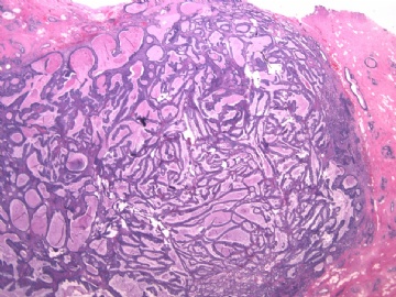
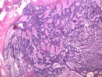
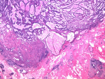
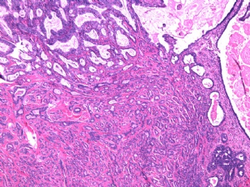
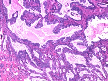
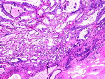
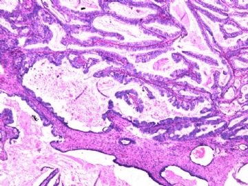
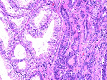
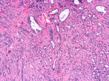
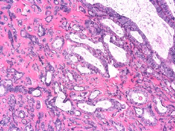
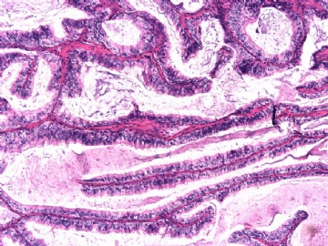
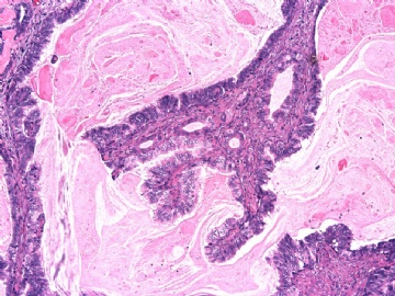
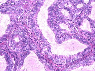
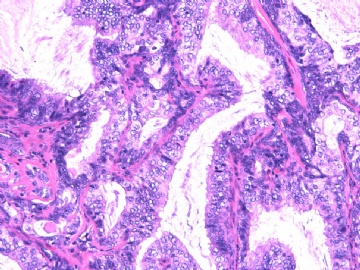
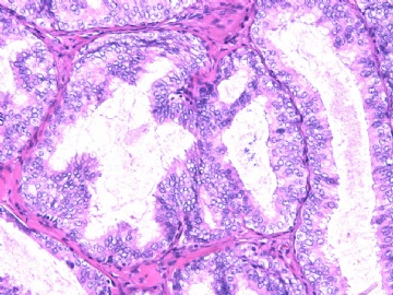
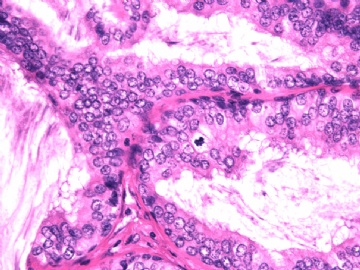
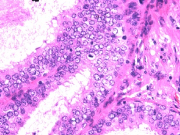
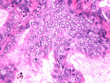
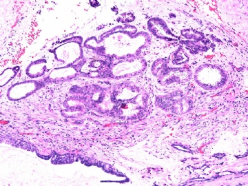
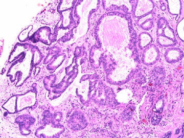
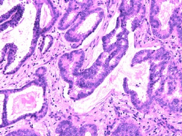
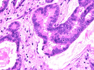
- B2892女性/35岁 左乳腺肿块3枚之一, 病理诊断?(IHC 2010-9-23)
| 姓 名: | ××× | 性别: | 年龄: | ||
| 标本名称: | 左乳腺肿块切除标本 | ||||
| 简要病史: | 发现左乳多发性肿块二周。 | ||||
| 肉眼检查: | 乳腺切除组织二块,其中见3个结节,其中一个1.5x1.2x1.0cm,边界清楚,部分有包膜,切面灰红色,无粉刺样物。 | ||||
其他二枚肿块为典型纤维腺瘤。
-
本帖最后由 于 2010-09-23 05:47:00 编辑

- xljin8
相关帖子
- • 有挑战性吗?
- • 乳腺肿物
- • 乳腺包块,诊断?
- • 乳腺肿物
- • 左乳肿物
- • 乳腺肿物一例
- • 腺病?癌?其他?(12楼常规,24楼免疫组化及会诊结果)
- • 求助:56岁女,左乳肿物,能排除小管癌吗?
- • 38岁乳腺(新加HE切片)
- • 乳腺肿块
-
本帖最后由 于 2010-09-23 05:43:00 编辑
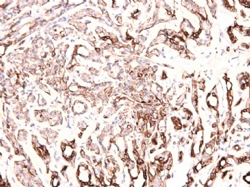
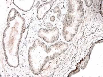
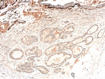
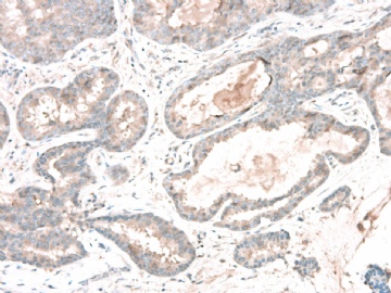
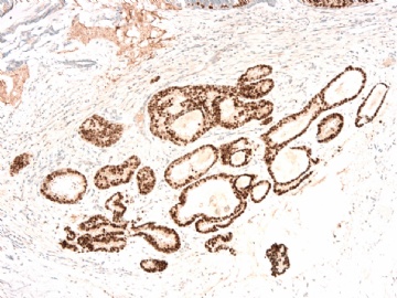
IHC 标记结果:
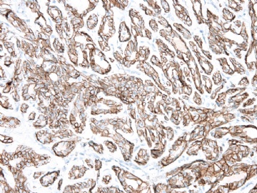
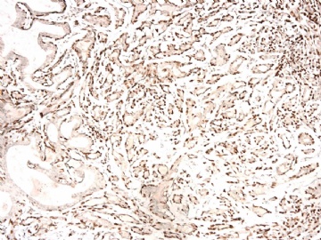
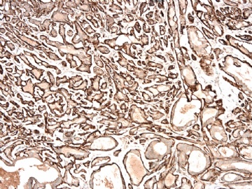
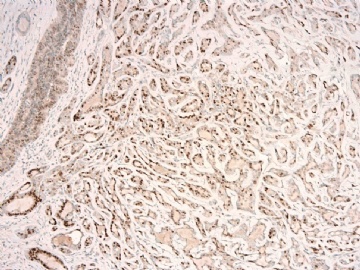
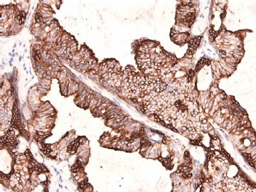
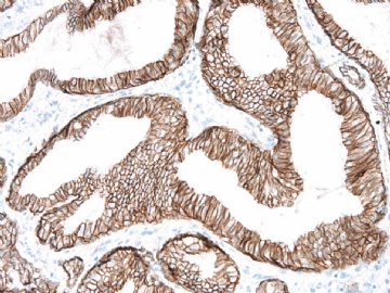
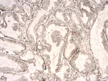
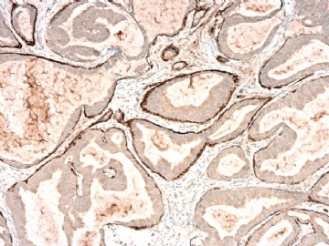
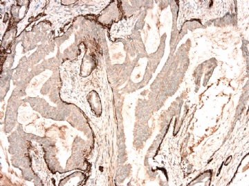
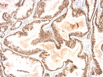
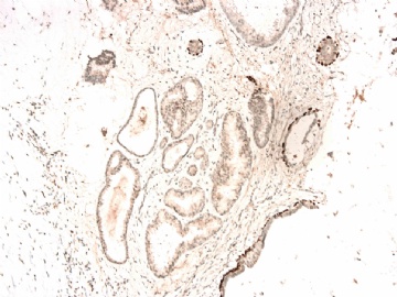
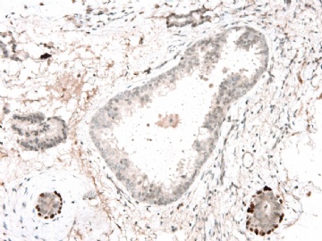
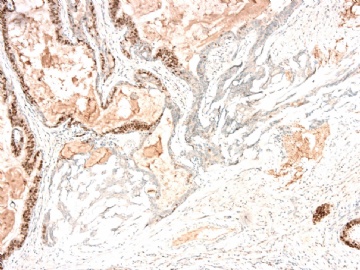
图1 34BE12、图2 E-cadherin、图3 P63、图4 SMMHC、 图5 ER、 图6 34BE12、图7 E-Cadherin、图8 P63、图9-10 SMMHC、 图11 ER、 图12-13 P63、 图14 P63、图15-16 SMMHC、 图17-18 ER。
请注意IHC图17左上角和图18右侧的形态,是在IHC标记片中新发现的形态学改变。

- xljin8
-
本帖最后由 于 2010-09-29 20:38:00 编辑
P63 stain shows this area may be invasive tumor, but i am not sure the invasion based on H&E. True glass slide reading can be helpful.(p63染色显示这个区域可能是浸润性肿瘤。但根据HE染色我不能确定浸润。看看切片可能有帮助。)
Sugggestion: (建议)
1. repeat SMMHC stain or add another myoepithelial marker, such calponin (重复SMMHC染色或者加做其它肌上皮标记,如calponin)
2. Submit more sections for microscopic examination.(补充取材制片观察)
As I mentioned before, our patients always had core biopsy before segmental mastectomy or excisional biopsy. If pathologic diagnoses are DCIS, ADH, papilloma, ALH et al, we need to submit all segmental mastectomy or excional biopsy specimen for microscopic examination. This is why 40-60 slides are very common for one case of excional bx or segmental mastectomy. The high number is 267 slides for one case of DCIS. I signed out breast large last week. I had 450 slides for one day and 500 slides for another day.(我以前提到,乳腺区段切除或切除活检之前,我们先做粗针穿刺活检。如果诊断了DCIS、ADH、乳头状瘤、ALH等,乳腺区段切除或切除活检全部标本都要取材。这就是乳腺区段切除或切除活检的取材量多达40-60张切片的原因。最多取过1例DCIS达267张切片。上周我签发了大量乳腺报告。一天有450张切片,另一天多达500张)
I do not think we can have routine daily practice as I mention above in China. I knew one case may be 40 RMB for one case. However for some special cases Submitting more sections are needed.
-
本帖最后由 于 2010-09-29 20:40:00 编辑
Dr. Jin's cases often are difficult and interesting. Thank for sharing.
Suggestion to Dr. Jin: Please send IHC photos separately in future if you can. Otherwise it is very difficult to figure out what stains they are.
(Dr.Jin的病例常常很难,也很有趣。谢谢分享!
建议Dr.Jin:以后可以把IHC图片分别上传。否则很难弄清楚,某图是什么免疫染色。)

