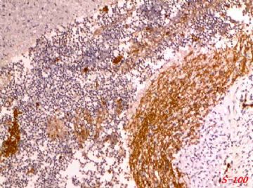
| 图片: | |
|---|---|
| 名称: | |
| 描述: | |
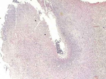
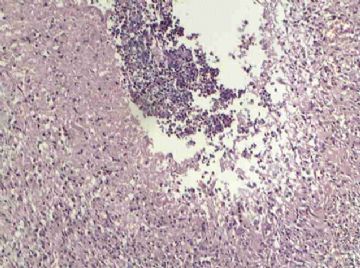
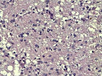
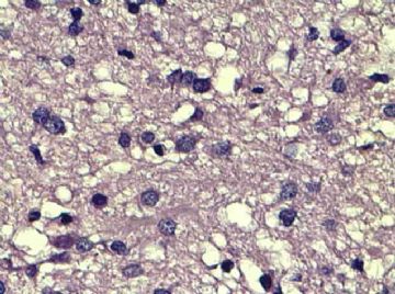
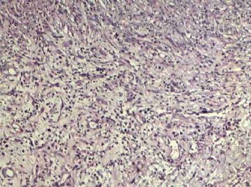
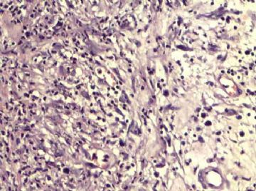
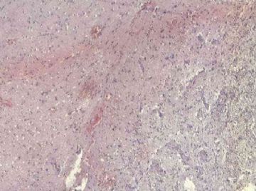
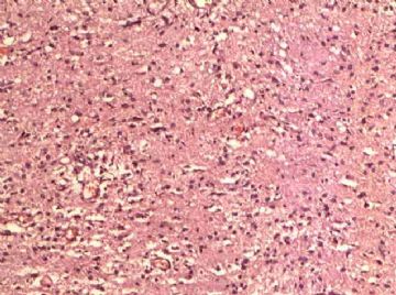
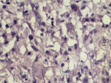
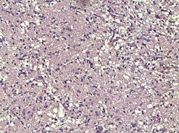
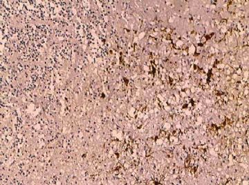
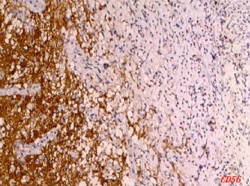
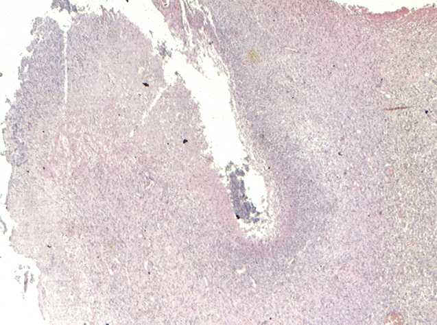
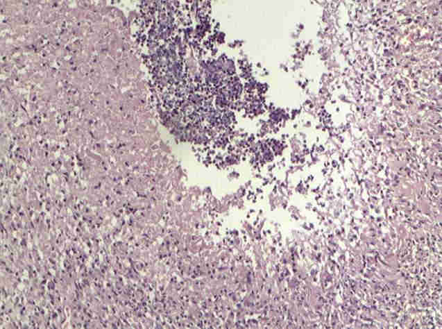
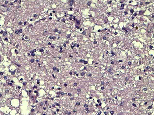
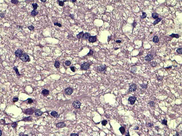
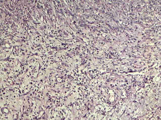
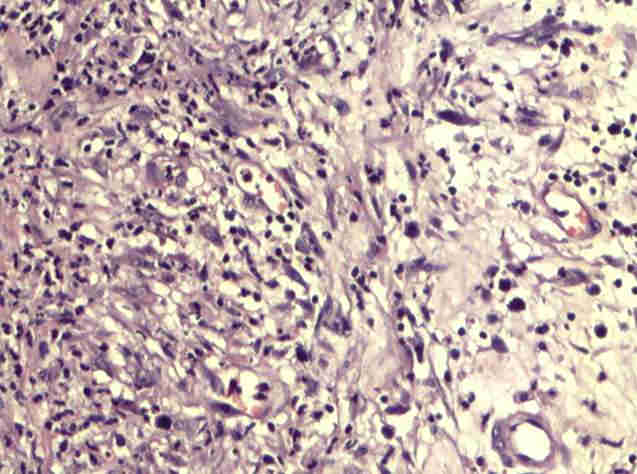
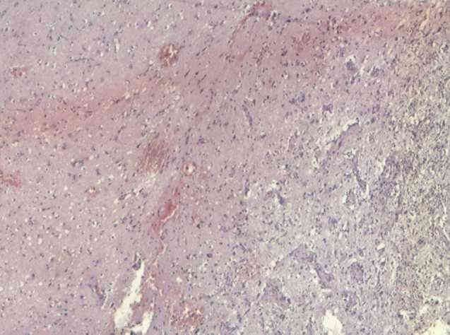
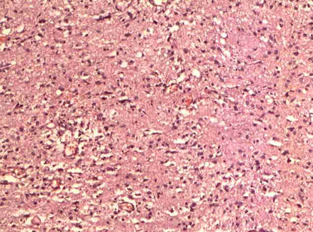
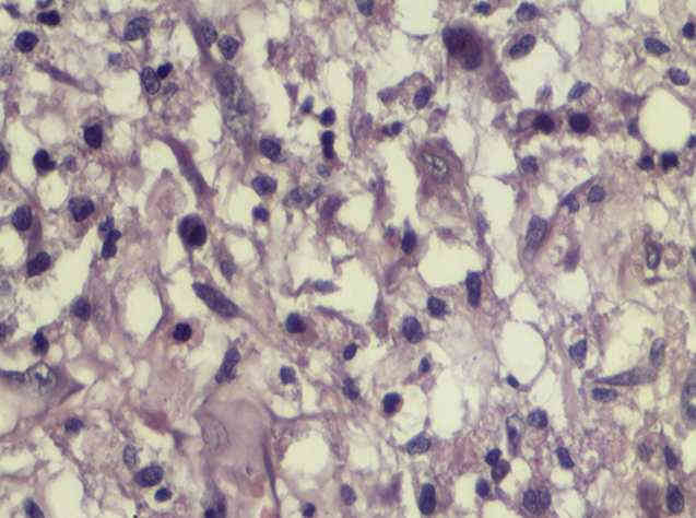
- 脑脓肿 不能排除胶质瘤?
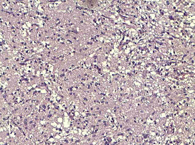
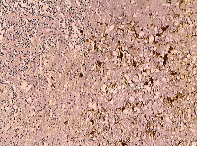
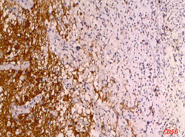
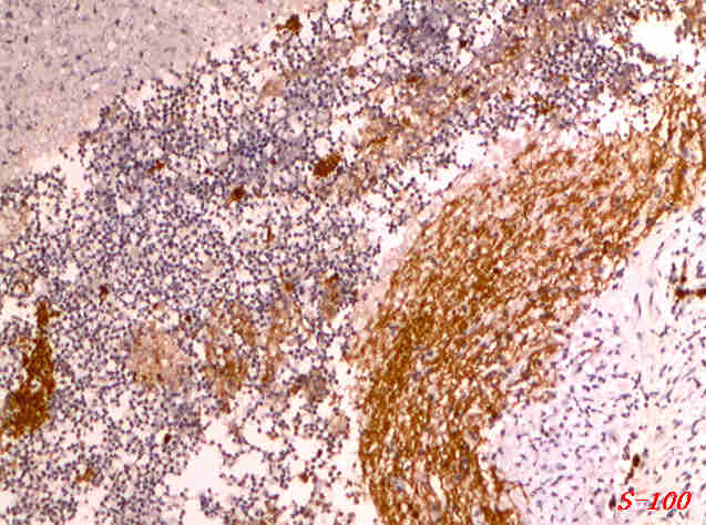
| 姓 名: | ××× | 性别: | 男 | 年龄: | 37岁 |
| 标本名称: | 左颞叶手术切除标本 | ||||
| 简要病史: | “ 头痛头晕9天,发现左颞”5天收入院.2010-7-10外院MR示:左颞叶占位:脓肿?不除外脑肿瘤。手术所见:嚢液黄白色。实验室检查:脓性。 | ||||
| 肉眼检查: | 灰白及灰红色囊状组织一块,V:6.5×4×3CM,切面可见一囊腔,长径约4CM,囊壁厚0.3-1.2CM。 | ||||
标签:
-
本帖最后由 于 2010-08-07 11:43:00 编辑
×参考诊断
| 以下是引用mjma在2010-8-7 1:52:00的发言: From the limited two photos shown so far, this is an abscess and not glioma. But more sampling of representative photos, in higher magnification, would make this interpretation certain. |
-
本帖最后由 于 2010-08-10 16:44:00 编辑
| 以下是引用fyshan在2010-8-10 4:41:00的发言:
脑脓肿 vs 胶质瘤 should be easily seperated by MRI scan. By pathology, 脑脓肿 has lots of fibroblast proliferation in 脑脓肿 wall and special stain, like reticulin or trichrom may be helpful.
Agree with Dr. Ma's analysis.
Good luck. |
