| 图片: | |
|---|---|
| 名称: | |
| 描述: | |
- 左顶枕叶肿瘤切除(P103119)
| 姓 名: | ××× | 性别: | 男 | 年龄: | 42 |
| 标本名称: | 左顶枕叶肿瘤 | ||||
| 简要病史: | 复视半年,头痛20天 | ||||
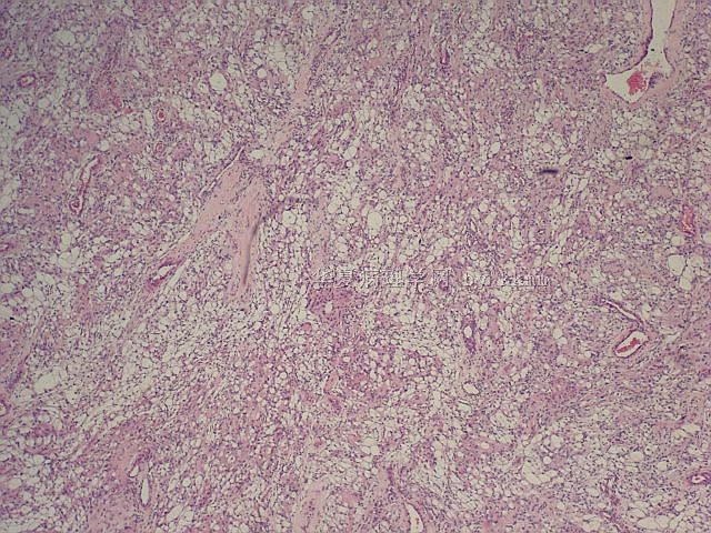
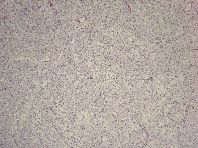
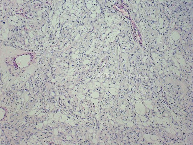
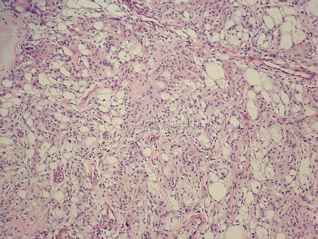
| 肉眼检查: | 左顶枕交界区镰窦旁质韧肿瘤,血供丰富 | ||||
名称:图1
描述:图1
名称:图2
描述:图2
名称:图3
描述:图3
名称:图4
描述:图4
名称:图5
描述:图5
名称:图6
描述:图6
名称:图7
描述:图7
名称:图8
描述:图8
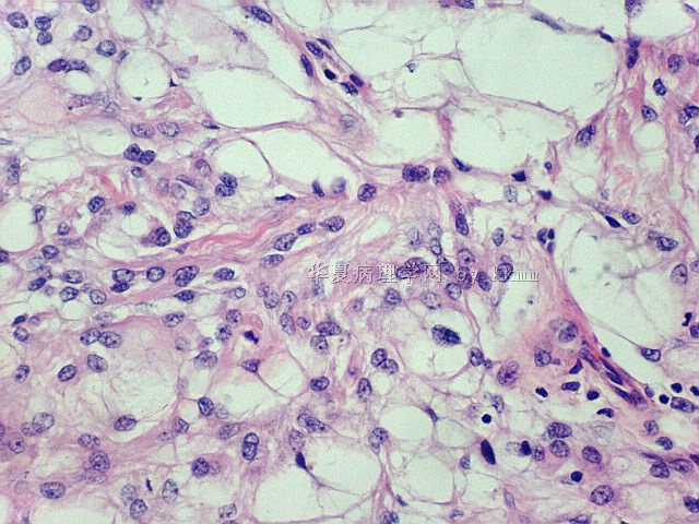
名称:图9
描述:图9
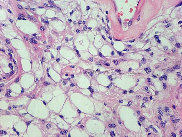
名称:图10
描述:图10
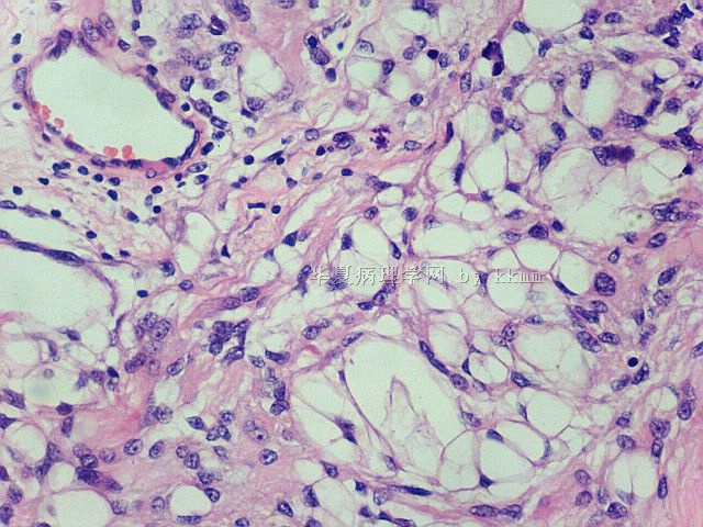
名称:图11
描述:图11
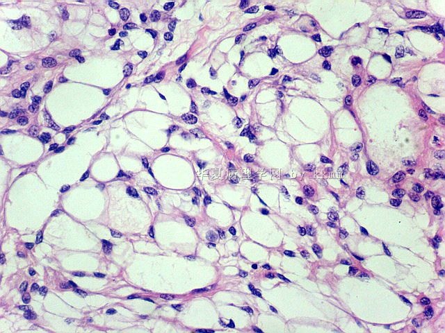
名称:图12
描述:图12
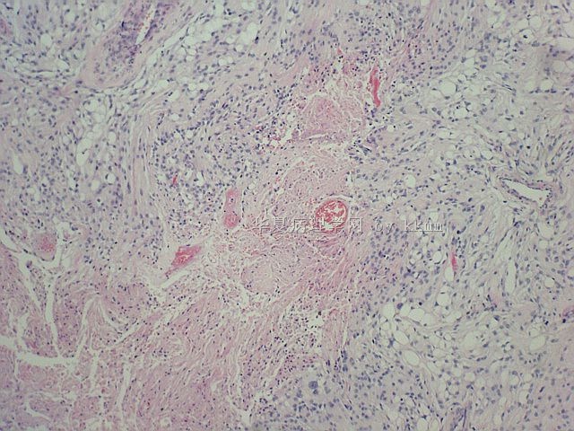
名称:图13
描述:图13
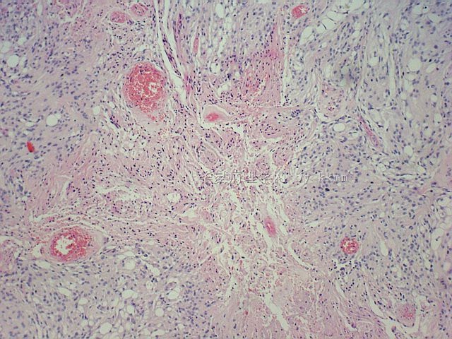
名称:图14
描述:图14
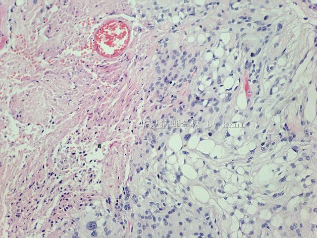
名称:图15
描述:图15
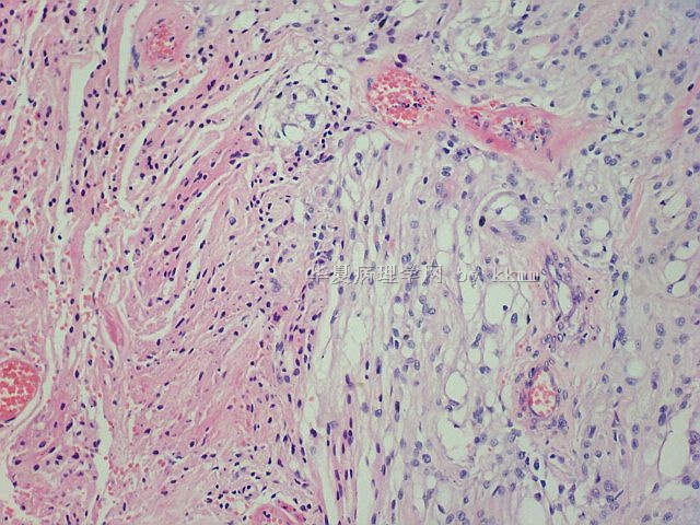
名称:图16
描述:图16
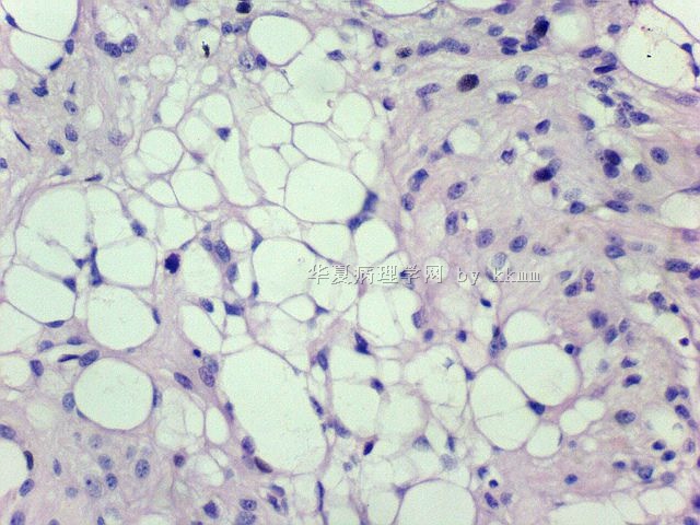
名称:图17
描述:图17
标签:
×参考诊断
-
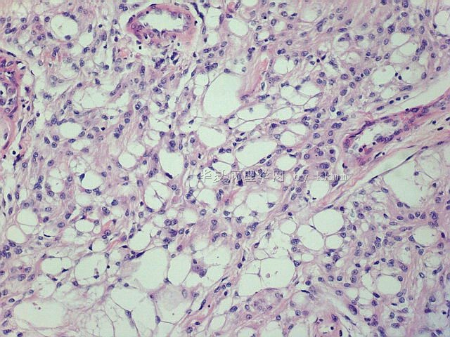
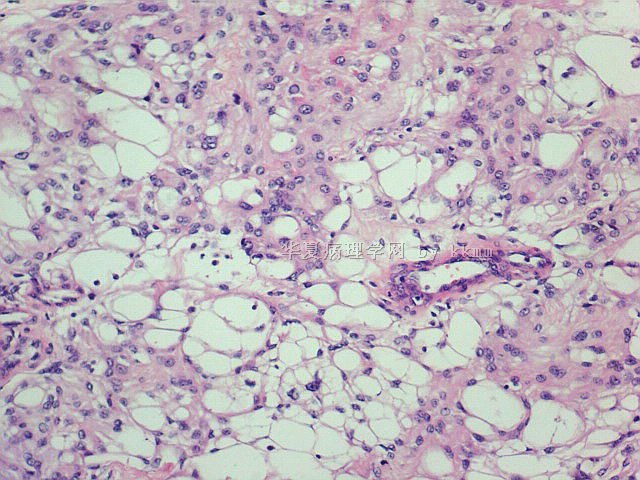
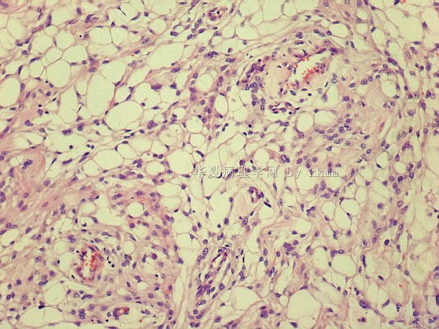
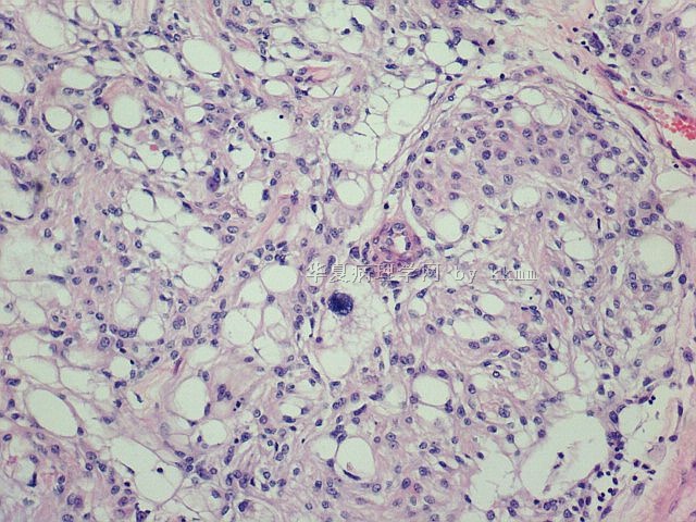
This is a case of WHO grade I meningioma of the microcystic type. Though focal tumor necrosis is seen, no other atypical features are sen. Careful mitotic counts have to be done, but from these photos I do not worry about atypical or malignant meningioma. This is not a case of clear cell meningioma, which is very rare and often found in younger adults. Nuclei of neoplastic cells in clear cell meningiomas are centrally located, not eccentric like in this case. The vacuolated cytoplasm in clear cell meningiomas is glycogen-rich (PAS-positive), but not that of microcystic meningioma.

聞道有先後,術業有專攻
| 以下是引用mjma在2010-7-29 8:51:00的发言: This is a case of WHO grade I meningioma of the microcystic type. Though focal tumor necrosis is seen, no other atypical features are sen. Careful mitotic counts have to be done, but from these photos I do not worry about worry about atypical or malignant meningioma. This is not a case of clear cell meningioma, which is very rare and often found in younger adults. Nuclei of neoplastic cells in clear cell meningiomas are centrally located, not eccentric like in this case. The vacuolated cytoplasm in clear cell meningiomas is glycogen-rich (PAS-positive), but not that of microcystic meningioma. |


