| 图片: | |
|---|---|
| 名称: | |
| 描述: | |
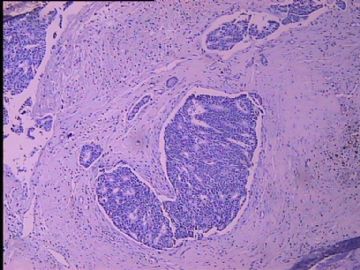
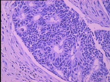
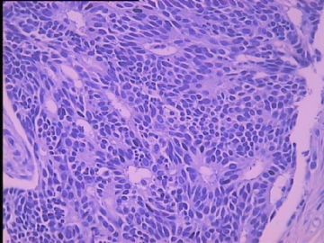
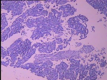
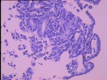
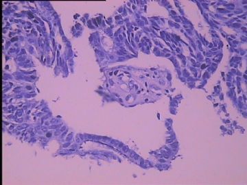
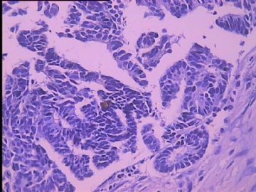
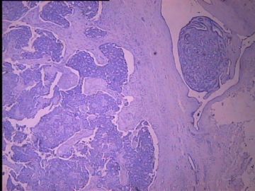
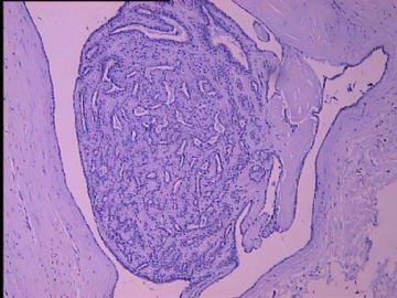
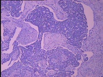
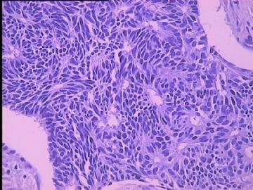
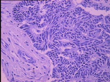
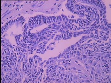
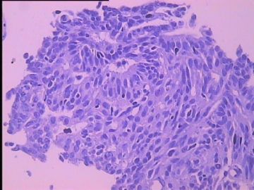
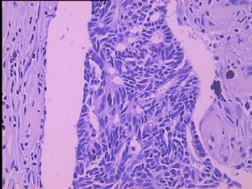
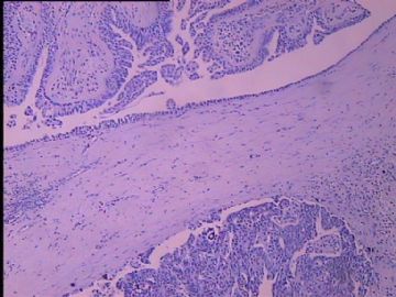
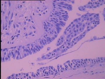
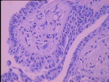
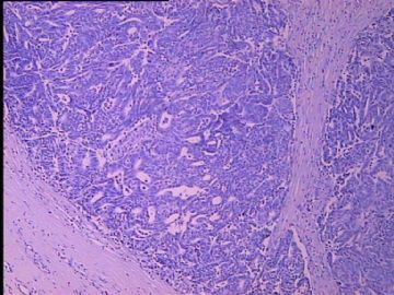
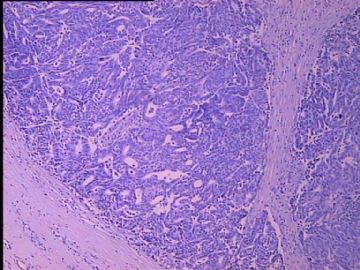
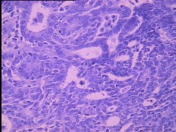
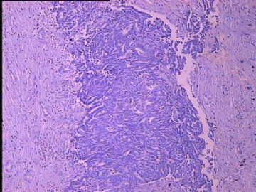
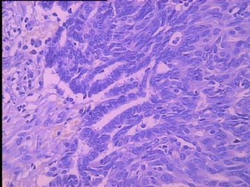
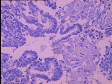
- B2752子宫内膜癌根治术后,乳腺肿物,请各位老师给看看
| 姓 名: | ××× | 性别: | 女 | 年龄: | 58 |
| 标本名称: | 乳腺肿物 | ||||
| 简要病史: | 子宫内膜癌根治术后,第四次化疗。发现右乳外下象限肿物一周。 | ||||
| 肉眼检查: | 不规侧组织一块,周边为脂肪组织,其内切面见肿物V2.0*1.5*1.5cm,肿物与周边切线界限尚清,切面暗红,质稍软,灰白,质中。 | ||||

- 把握今天,展望明天
相关帖子
Agree with above.
Need to rule out secondary tumor first. You can find the previous slides of endometrial ca and compare the morphology with breast lesions.
Feeling liker a papillary lesion of breast. Need to do myoepithelial markers to evaluate the nature of the lesions (benign, atypical, cancer) and to see if invasion is present.
Thank you for sharing this case.
-
本帖最后由 于 2010-07-02 11:59:00 编辑
| 以下是引用cqzhao在2010-6-26 18:01:00的发言:
Agree with above. Need to rule out secondary tumor first. You can find the previous slides of endometrial ca and compare the morphology with breast lesions. Feeling liker a papillary lesion of breast. Need to do myoepithelial markers to evaluate the nature of the lesions (benign, atypical, cancer) and to see if invasion is present. Thank you for sharing this case. |
赵老师的意见:
该病例首先要鉴别除外继发性肿瘤。复习原来宫内膜癌切片,并与当前的乳腺病变进行比较。
觉得本例很像是乳腺乳头状病变。因此需要作肌上皮标记来评价病变性质(良性、非典型性、癌?),还要看是否有浸润。

- 王军臣
