| 图片: | |
|---|---|
| 名称: | |
| 描述: | |
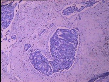
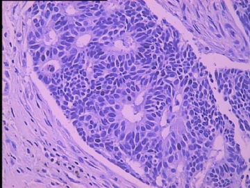
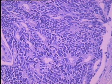
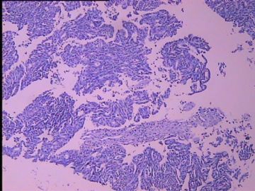
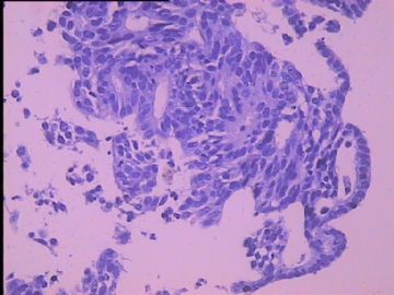
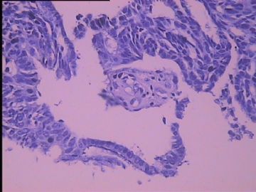
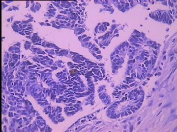
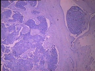
- B2752子宫内膜癌根治术后,乳腺肿物,请各位老师给看看
| 姓 名: | ××× | 性别: | 女 | 年龄: | 58 |
| 标本名称: | 乳腺肿物 | ||||
| 简要病史: | 子宫内膜癌根治术后,第四次化疗。发现右乳外下象限肿物一周。 | ||||
| 肉眼检查: | 不规侧组织一块,周边为脂肪组织,其内切面见肿物V2.0*1.5*1.5cm,肿物与周边切线界限尚清,切面暗红,质稍软,灰白,质中。 | ||||
标签:乳腺浸润性导管癌 导管内乳头状肿瘤

- 把握今天,展望明天
相关帖子
×参考诊断
浸润性导管癌,有内膜癌病史
-
CHENYINQIAO 离线
- 帖子:547
- 粉蓝豆:0
- 经验:593
- 注册时间:2010-07-20
- 加关注 | 发消息
| 以下是引用快乐之星在2010-8-22 19:39:00的发言:
请上级医院会诊,结果:浸润性导管癌. 免疫组化:ER+ PR± C-erbB-2 + Ki-67 3%+ p63- CD56- Syn- CD10- |
ER/PR cannot tell breast from endometrial origins.
P63- myoepithelial markers neg supports invasion ca. But need to see the photos to evaluate.
Do not know why stains for cd56 and syn were performed.
The patient can have invasive breast ca; DCIS and also papilloma. Cannot make definite dx based on the photos only.
Thank for sharing us the photos and data.

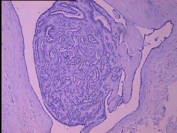
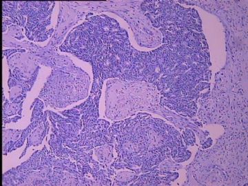
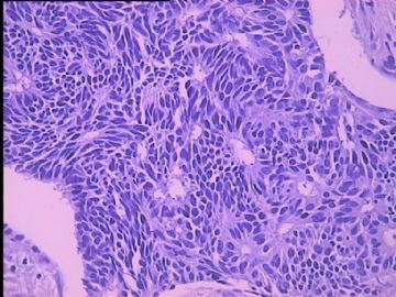
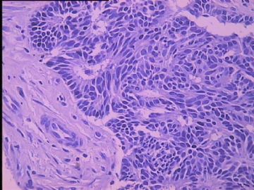

 愁死我了!不能强求病号去会诊,我作为一个“小兵”又不能擅自做主。只能找机会拿到大医院让专家给看看了。


