


| 图片: | |
|---|---|
| 名称: | |
| 描述: | |
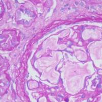
- 甲状腺乳头状癌?够诊断标准吗?
-
shihong4699 离线
- 帖子:1024
- 粉蓝豆:43
- 经验:2917
- 注册时间:2009-01-20
- 加关注 | 发消息
| 以下是引用zhanghx在2010-6-14 17:46:00的发言: 甲状腺乳头状癌?够诊断标准吗? |
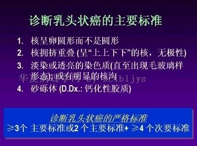
香港大学病理系陈国璋教授提出的甲状腺乳头状癌的诊断标准:
Major criteria for diagnosis of PTC
a.Nuclei ovoid rather than round
b.Crowded overlapping nuclei (manifesting as 'up and down' nuclei with no polarity)
c.Pale or clear chromatin (up to ground-glass appearance); or prominent nuclear grooving
d.Psammoma bodies (D.Dx.: calcified colloid
Minor criteria for diagnosis of PTC
a. Abortive papillae
b. Predominantly elongated or irregular-shaped follicles
c. Dark-staining colloid
d. Presence of nuclear pseudoinclusions
e. Multinucleated histiocytes in lumens of follicles
STRICT CRITERIA FOR DIAGNOSIS OF PTC
≥3 major criteria or 2 major criteria + ≥4 minor criteria

- 许春雷
-
WHO2004关于乳头状癌的描述,我的理解是:
1.乳头状癌的诊断依据主要是特征性的核的改变,虽然其组织结构在不同亚型间差别很大;
2.“在没有复杂乳头结构的肿瘤,乳头状癌的诊断则依赖这些核的特征,但必须在肿瘤中占据相当大的部分。”此点可以帮助我们区别乳头状癌和偶见乳头状癌核的特征的病变:如毒性甲状腺肿、慢性甲状腺炎、桥本甲状腺炎及某些滤泡性腺瘤等。但并没有提及可以根据“复杂乳头结构”即可诊断癌而不考虑核的改变的。
3.“癌性乳头应与结节性甲状腺肿的乳头状结构或伴乳头的滤泡性腺瘤相鉴别,也要与弥漫性增生中短的乳头状内折叠鉴别,后者的核是典型的圆形,位于基底部,而最重要的是缺乏乳头状癌核的特征。”换言之,复杂的乳头状结构必须有核的特征方可诊断癌。
4.“柱状细胞性乳头状癌,通常不具有典型乳头状癌的核特征...”此点可以作为普遍现象中存在的特殊现象来看待。
5.2004年WHO的诊断标准是目前最新的,代表了对乳头状癌研究的最新进展和认识,我觉得应以此为新的诊断标准,但暂时还存在不同的看法也是可以理解的。
- 许春雷
-
本帖最后由 于 2010-06-14 22:23:00 编辑
以下是引用xclbljys在2010-6-14 20:12:00的发言:
香港大学病理系陈国璋教授提出的甲状腺乳头状癌的诊断标准: Major criteria for diagnosis of PTC 2009年出版的Modern Surgical Pathology第二版中有关甲状腺乳头状癌的诊断标准如下: Diagnostic Criteria for Papillary Carcinoma Based on a constellation of features, no single one of which is pathognomonic. For a noninvasive tumor, the diagnostic label papillary carcinoma should be applied only when the typical cytologic features are well developed. Basic Criteria 1. Cytologic features: ovoid nuclei that are crowded, without polarization, clear or pale, and grooved, with or without pseudoinclusions 2. demonstration of vascular or capsular invasion not required Strong Supporting Feature, if Present 1. Psammoma bodies Other Supporting Histologic Features 1. Papillae (including abortive papillae) 2. Follicles that are elongated or irregularly shaped 3. dark-staining colloid 4. multinucleated histiocytes in lumens of follicles or papillae Deceptively “Benign” Patterns Warranting Serious Consideration of the Possibility of Papillary Carcinoma 1. “Colloid nodule” with delicate papillary budding in some follicles, or psammoma bodies, or many clear nuclei (papillary carcinoma, macrofollicular variant) 2. Picture resembling Hashimoto’s thyroiditis or lymphocytic thyroiditis, but with many “knife marks” on the histologic section because of the presence of psammoma bodies (diffuse sclerosing variant of papillary carcinoma) 3. “Follicular adenoma” with many elongated follicles and dark-staining colloid or abortive papillae (papillary carcinoma, encapsulated follicular variant) 4. “Degenerate cyst” but with occasional small papillary tufts projecting into the lumen or some follicles in the fibrous wall lined by cells with high nuclear-to-cytoplasmic ratio (cystic variant of papillary carcinoma) 5. Spindle cell proliferation resembling nodular fasciitis or fibromatosis (papillary carcinoma variant with exuberant nodular fasciitis–like stroma) 该书中甲状腺和甲状旁腺一章的作者是: John K. C. Chan, MD Consultant Pathologist, Institute of Pathology, Queen Eliza- beth Hospital, Kowloon, Hong Kong, China 与9楼提及的香港大学病理系陈国璋教授是不是同一作者? |
不得不说了
1。 xclbljys 老师所说的“标准”已经过时了,包括其原始提出者陈国璋教授,也已经不再提所谓的几加几了
2。zhanghx老师提到的是09年的Modern Surgical Pathology,其中的甲状腺部分就是陈国璋教授所写,由此可以看出陈教授的观点。说是改变也好,说是新“标准”也好(其实病理并无标准可言),可以看出,甲状腺乳头状癌的诊断,重点在“核特征”。至于核特征到底有多少条?是不是还得满足几条才够?我认为不必争吵,够了的一眼就够。其关键在于:与正常的进行对比。
3。其实Modern Surgical Pathology中,关于甲状腺乳头状癌的前面有一段,或许意义更大:Papillary carcinoma is defined as a malignant epithelial tumor showing evidence of follicular cell differentiation and characterized by distinctive nuclear features. In other words ,the key to diagnosis lies in the nuclear characteristics, and the demonstration of invasive growth is not required.
- 赚点散碎银子养家,乐呵呵的穿衣吃饭
谈不到请教,大家共同学习
无限玄机:确实是这样
核特征:
阿克曼中讲:毛玻璃样核,核内假包涵体,核沟,核内微丝
Nikiforov YE等人最新著作中提到:核增大、排列拥挤重叠、轮廓不规则、核沟、假包涵体、毛玻璃样核
Boerner SL等人还提出了:显著的核仁
其实仔细想想:乳头状癌,首先是肿瘤,而肿瘤的特性是不受限制的增殖。其特征性表现之一就是核增大,这是基础,导致了拥挤重叠、导致了层次增多。这也就是我上面提到的“对比”的意义之所在:只有对比,才能知道是不是大了……
以上文字,来自前辈无数次的教导,还有很多没有体现出来,水平有限,抱歉……
- 赚点散碎银子养家,乐呵呵的穿衣吃饭
-
本帖最后由 于 2010-06-15 15:17:00 编辑
| 以下是引用wq_9603在2010-6-15 0:16:00的发言:
谈不到请教,大家共同学习 无限玄机:确实是这样 核特征: 阿克曼中讲:毛玻璃样核,核内假包涵体,核沟,核内微丝 Nikiforov YE等人最新著作中提到:核增大、排列拥挤重叠、轮廓不规则、核沟、假包涵体、毛玻璃样核 Boerner SL等人还提出了:显著的核仁 其实仔细想想:乳头状癌,首先是肿瘤,而肿瘤的特性是不受限制的增殖。其特征性表现之一就是核增大,这是基础,导致了拥挤重叠、导致了层次增多。这也就是我上面提到的“对比”的意义之所在:只有对比,才能知道是不是大了…… 以上文字,来自前辈无数次的教导,还有很多没有体现出来,水平有限,抱歉…… |
听君一席话,胜读十年书!受益匪浅!请结合您的体现谈一下您对本例的诊断意见,最好有诊断依据!
也希望zhangHY网友发表诊断意见!谢谢!
- 许春雷
-
以下是引用xclbljys在2010-6-14 20:18:00的发言:
2004年WHO的诊断标准是目 前最新的,代表了对乳头状癌研究的最新进展和认识,我觉得应以此为新的诊断标准,但暂时还存在不同的看法也是可以理解的。
2007年版的Diagnostic Histopathology of Tumors和Diagnostic Criteria Handbook in Histopathology,以及2009年版的Modern Surgical Pathology这三部著作中提及甲状腺乳头状癌的诊断标准,2004版的WHO Classification of Tumours一书中何处讲到甲状腺乳头状癌的诊断标准?请赐教!
-
本帖最后由 于 2010-06-15 17:51:00 编辑
| 2004版的WHO Classification of Tumours一书中何处讲到甲状腺乳头状癌的诊断标准?请赐教! |
问的好
WHO的著作写明了“WHO classification of tumor"
而不是criteria
就其内容而言,也是“Histopathology”
其第一句话就是“The characteristic nuclear features of papillary carcinoma include enlargement, oval shape ……”
哪有啥标准之说呢
真的有了如同血常规一样的标准就好了
我们也可以轻松了
- 赚点散碎银子养家,乐呵呵的穿衣吃饭


