| 图片: | |
|---|---|
| 名称: | |
| 描述: | |
- 第一次试发贴:胃溃疡活检
-
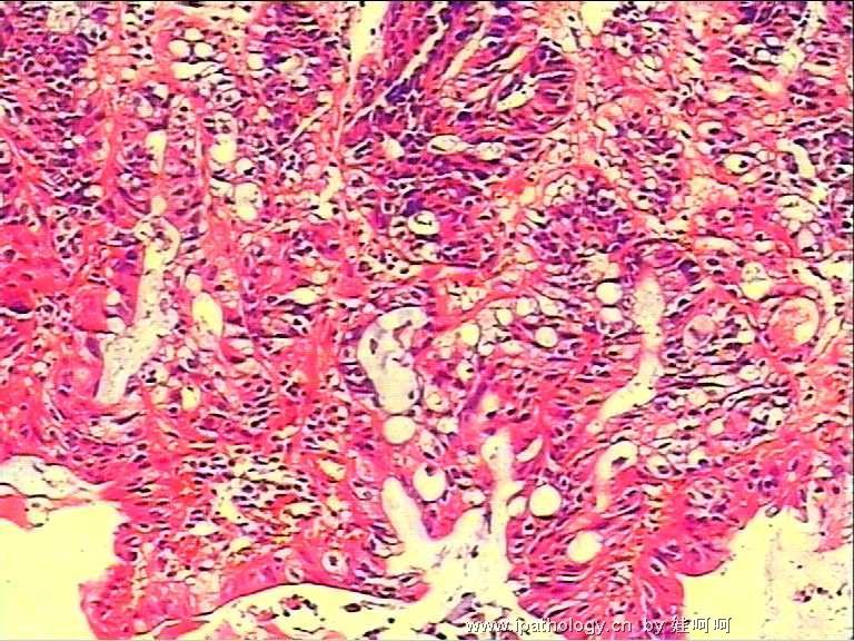
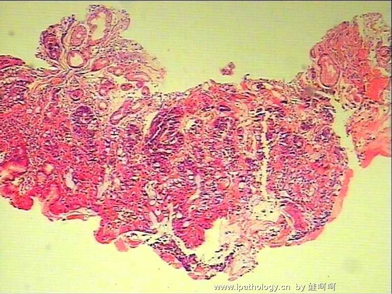
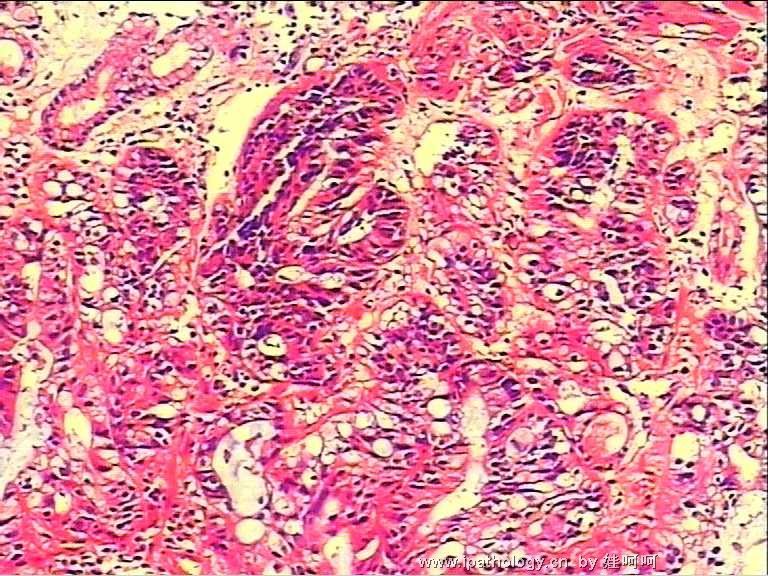
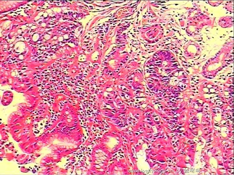
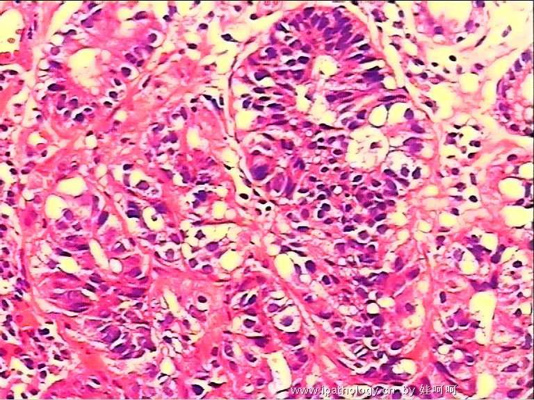
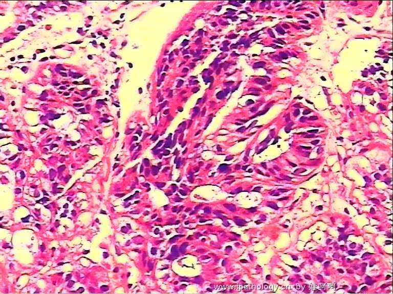
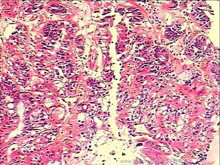
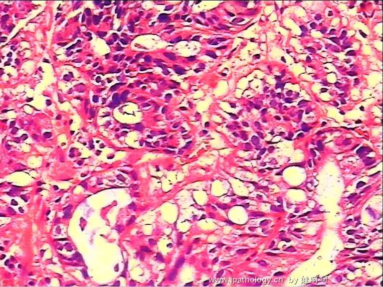
The histology seems suboptimal from the photos. The glandular lining and glandular architecture of this mucosal biopsy appear very atypical. Many glandular lining epithelia are crowded, have hyperchromatic nuclei and vacuolated cytoplasm. However, the surface lining epithelia are normal without apparent intestinal metaplasia. These features raise two important differential diagnoses - (1) severe epithelial dysplasia or adenocarcinoma in situ with signet ring cell formation, and (2) neuroendocrine cell hyperplasia as part of ulcer repair. I have not found isolated signet ring cells in the lamina propria, but I would be very careful to be sure of this point under the microscope. Therefore I would request histology to recut a few deeper sections for evaluation. If isolated signet ring cells are seen in lamina propria, this case is signet ring cell poorly differentiated adenocarcinoma. If not, I at least get to see more of these atypical cells, hopefully with better histologic details. If histology cannot be improved and things remain the same as shown here, I would consider using immunohistochemistry (including p53, chromogranin A, synaptophysin, NSE, and AE1) to further delineate these cells in question or simply recommend re-biopsy of the lesion to be certain of its nature.

聞道有先後,術業有專攻
-
mjma,would you answer me some questions?
1what's the differences between atypical hyperplsia and dysplasia?
2if the stains of cgA ,SYN,NSE are positive,how do you tell neuroendocrine cell hyperplasia
from neuroendocrine cell neoplasma?
3what's the standard when you diagnose signet ring cell carcinoma?
4what's the standard do you use to divide epithelial dysplasia into low-grade and high grade?
Thank you very much!


| 以下是引用忍者神龟 在2006-10-25 23:20:00的发言: 1. what's the differences between atypical hyperplsia and dysplasia? |
2. Neuroendocrine cell hyperplasia in the gastric mucosa is common in certain conditions (such as atrophic gastritis of autoimmune etiology), and eventially will give rise to carcinoid tumor, which in turns may progress to a neuroendocrine carcinoma when there is stromal invasion and sufficient cytologic atypia.
3. Signet ring cell carcinoma of the gastrointestinal tract has to show isolated or small groups of atypical cells with eccentric nuclei and large cytoplasmic mucinous vacuoles in the stroma.
4. Epithelial dysplasia in gastrointestinal tract mucosa is mostly divided into high and low grade. Most small adenomatous polyps of colonic and rectal mucosae show only low-grade dysplasia - nuclei stay near the basal aspect of the dysplastic cells. To diagnose high-grade dysplasia (including adenocarcinoma in situ), the nuclei have to be stratified and cover all aspects of the columnar cells, and nuclear size and shape are much more variable.

聞道有先後,術業有專攻
-
学习mjma老师的讲解并试译如下:
第2楼
图片质量不佳。此例粘膜活检中,腺体排列和腺结构显得很不典型。许多腺上皮受挤压,核染色质深染,胞浆内有空泡。然而表面被覆上皮是正常的,没有明显的肠化生。这些特征提示两个重要的鉴别诊断:(1)重度上皮异型性或原位腺癌伴印戒细胞形成;(2)神经内分泌细胞增生,这是溃疡修复的表现之一。我没有发现孤立的印戒细胞存在于固有层之中,但是,如果是在显微镜下,我必须非常小心地明确这一点。因此我可能会要求重切片、深切以作进一步评价。如果不这样,我至少要见到更多的这些不典型细胞,以图发现更好的组织学细节。如果切片质量不能提高并且仍像这些图中所见,我会考虑免疫组化(包括p53,CgA,Syn,NSE,AE1)进一步显示这些有疑问的细胞,或者只是建议病灶处再次活检以明确病变性质。
第10楼
以下是引用忍者神龟的发言:
1.不典型增生和异型增生的区别?
2.如果CgA,SYN,NSE阳性,如果区分神经内分泌细胞增生或其肿瘤?
3.你诊断印戒细胞癌的标准?
4.你区分上皮异型的低/高级标准?
mjma老师的回答:
1、不典型增生是良性、反应性过程,异型增生是真正的癌前改变。
2、胃粘膜内神经内分泌细胞增生在某些情形下是常见的(如:自身免疫导致的萎缩性胃炎),并且最终会导致类癌之类的肿瘤;进而,当存在间质浸润和足够的细胞学不典型性时,可能进展为神经内分泌癌。
3、胃肠道印戒细胞必须显示出现在间质内的、孤立的或小灶性不典型细胞,伴偏心核和大的胞浆粘液空泡。
4、胃肠道粘膜的上皮异型增生通常分为高、低级别。结直肠粘膜的大多数小的腺瘤性息肉只显示低级别异型增生——异型性核仅见于下层。诊断高级别异型增生(包括原位癌)要求核复层化、全层显示异型性、核大小和形状变化更大。

华夏病理/粉蓝医疗
为基层医院病理科提供全面解决方案,
努力让人人享有便捷准确可靠的病理诊断服务。

