| 图片: | |
|---|---|
| 名称: | |
| 描述: | |
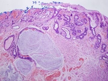
- 谈东风病例9 Case T0009:食道病变, 有浸润吗? If yes, How deep?
| 姓 名: | ××× | 性别: | 58 | 年龄: | male |
| 标本名称: | |||||
| 简要病史: | Recent dysphasia. | ||||
| 肉眼检查: | 1.2cm lesion in the middle portion of esophagus. | ||||
How deep is the lesion??
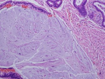
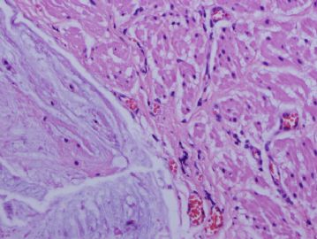
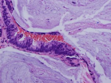
Diagnosis: focal early adenocarcinoma within the muscularis mucosae (病变应是位于黏膜肌层).
This outside case was called as an invasive adencarcinoma involving the muscularis propria (肌层).
1. The muscularis mucosae in the esophagus, particularly in the lower middle and distal parts of esophagus, can be very thick. In the Barrett's esophagus, the muscularis mucosae would be "double layer". It is common error to call this muscularis mucosae as muscularis propria. But the tumor stage is significantly different.
2. Adenocarcinoma of the esophagus is very common in the western countries. For example, adenocarcinoma of the esophagus in U.S.A. is much more common than adenocarcinoma of the stomach. Most adenocarcinomas in the esophagus arise in a background of Barrett's esophagus (also see the second case in 第 14 楼). The criteria to call early adenocarcinoma is mainly based on a) archiectural disorder and b)early invasiveness, namely, the neoplastic glands penetrate through the basement membrane into the muscularis mucosae. Sometimes, the changes is very subtle, like this case, in which only one gland shows malignant transformation. The case in is more obvious, like the case in 第 14 楼.
-
本帖最后由 于 2010-06-06 02:19:00 编辑


