


| 图片: | |
|---|---|
| 名称: | |
| 描述: | |
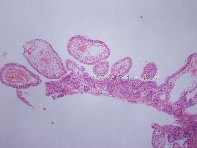
- 谈东风病例6 Case T0006 62岁女性, 下腹痛10周,十二指肠活检
 图1
图1 图2
图2 图3
图3 图4
图4 图5
图5
| 姓 名: | ××× | 性别: | female | 年龄: | 62 |
| 标本名称: | 十二指肠活检 | ||||
| 简要病史: | 62岁女性, 下腹痛10周 and watery stools (5-6 times a day) for 10 weeks. Clinically, it was suspicious for infection since the patient recently traveled to Mexico. | ||||
| 肉眼检查: | Endoscopically, the mucosa of the duodenum and terminal ileum shows edema-like appearance. | ||||
This is a rare case, a vice Minister with a lymphoma presenting as an initial GI involvement. The whole story is following:
This is a 62-year-old woman who began having diarrhea (up to 6 times per day, very loose, with no definite blood)for 3-4 months. The endoscope examination showed duodenum and terminal ileum with edema-like hypertrophic villi/mucosa in a patchy distribution.
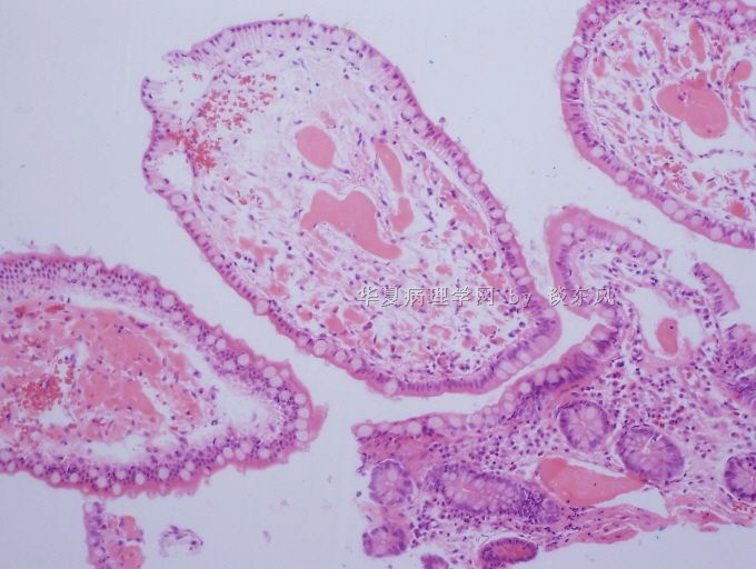
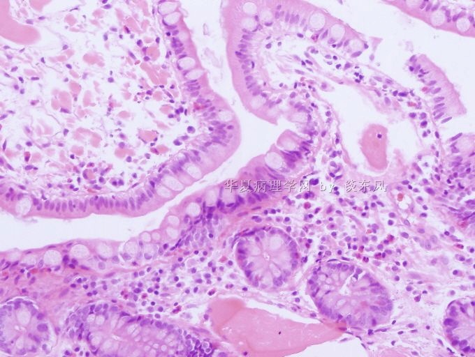
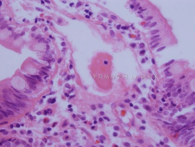
Biopsy shows villi are distended by amorphous eosinophilic hyalinized material. Notice, some of the hyalinized material looked like in the lymphatic spaces, but 1) the color of the material is much dense and dark than lymphoid fluid, and 2) the material is associated with lymphocytes and plasma cells. Initial study included Congo red stain and PAS stain. Congo red was negative. PAS stain was positive. Subsequently, IgA and IgM immunostains were performed. IgM was strongly positive (shown in Figure 5 below).
Diagnosis: IgM gammopathy involving intestine. Recommend clinical work-out for lymphoid proliferative disorders, including plasmacytoid lymphoma(Waldenstrom macroglobulinemia), lymphoplasmacytic lymphoma, among others.
Subsequently, bone marrow biopsy was performed. It showed 1) marked increase of lymphocytes (numerous small and intermediate to rare large size lymphoid cells with moderate to abundant amounts of pale, agranular cytoplasm. The nuclear morphology ranges from round to ovoid with evenly distributed condensed, mature appearing chromatin. Nucleoli are absent), and 2) marked increase of plasma cells ( display a spectrum of morphologies ranging from typical to slightly enlarged cell size with nucleomegaly, increased N:C ratios and indistinct nucleoli. Occasional plasma cells demonstrate clear cytoplasmic vacuoles).
Flow cytometric immunophenotypic studies were performed on portions of the bone marrow aspirate identified a monotypic kappa B-lymphoid and plasma cell population. The B-cells are positive for CD19 and CD20, but are negative for CD10. A subpopulation appears to have variable CD5 expression. The monotypic plasma cells are positive for CD19, CD38, CD52, CD138 and are negative for CD20 and CD56. These findings support a lymphoplasmacytic neoplasm.
FINAL DIAGNOSIS
LYMPHOPLASMACYTIC LYMPHOMA INVOLVES BONE MARROW AND INTESTINE.
标签:
-
本帖最后由 于 2010-06-02 01:16:00 编辑
×参考诊断
discussion and a new photo(Fig.5) below.
| 以下是引用谈东风在2010-5-11 6:18:00的发言:
More photos.
Notice that the dark dense pink material is mixed with cells. The pink material is within the laminar propria. The details of the cells are shown in the last photo. |
The dense pink material is thicker and darker than lymph fluid. The mixed cells are predominately plasma cells.


