| 图片: | |
|---|---|
| 名称: | |
| 描述: | |
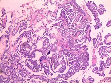
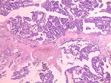
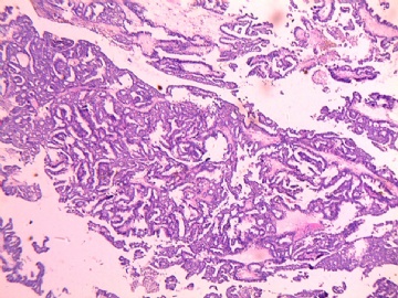
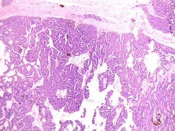
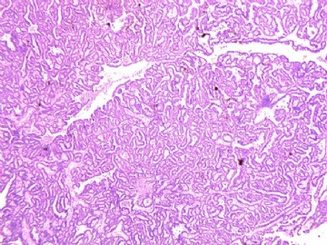
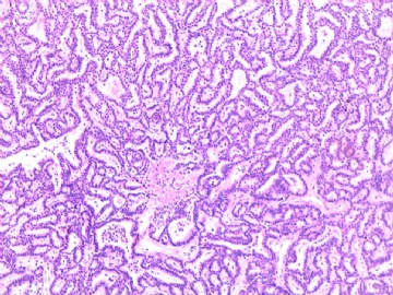
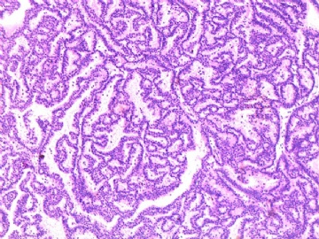
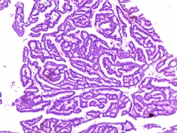
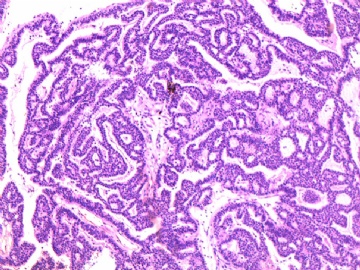
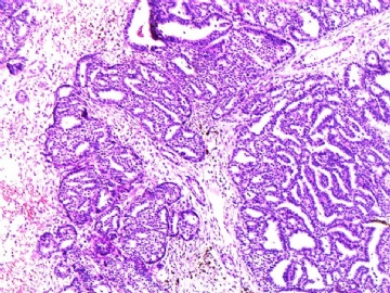
- B2674左侧乳腺肿瘤
| 姓 名: | ××× | 性别: | 年龄: | 65 | |
| 标本名称: | |||||
| 简要病史: | 左乳肿块5年 | ||||
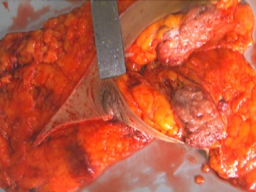
| 肉眼检查: | 大约5cm,灰红色,有出血,边界不清。无明显囊壁。 | ||||
-
本帖最后由 于 2010-05-07 11:04:00 编辑
相关帖子
http://www.ipathology.com.cn/forum/forum_display.asp?keyno=106223
http://www.ipathology.com.cn/forum/forum_display.asp?keyno=106922
For some ones who are intersted to these papillary lesions, please check above links with some cases and discussion.
-
xjdong2009 离线
- 帖子:6
- 粉蓝豆:1
- 经验:6
- 注册时间:2008-12-30
- 加关注 | 发消息
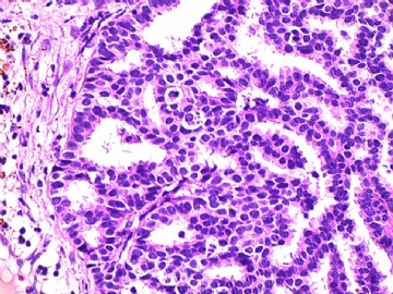
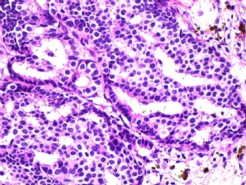
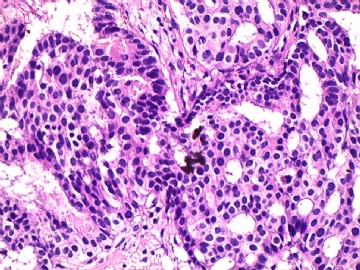
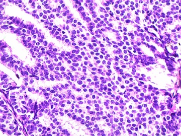
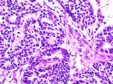
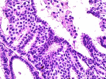
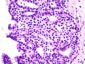
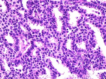
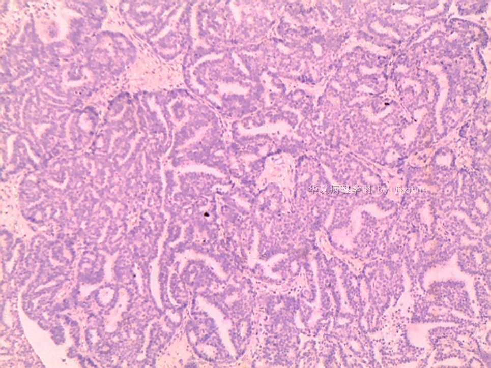
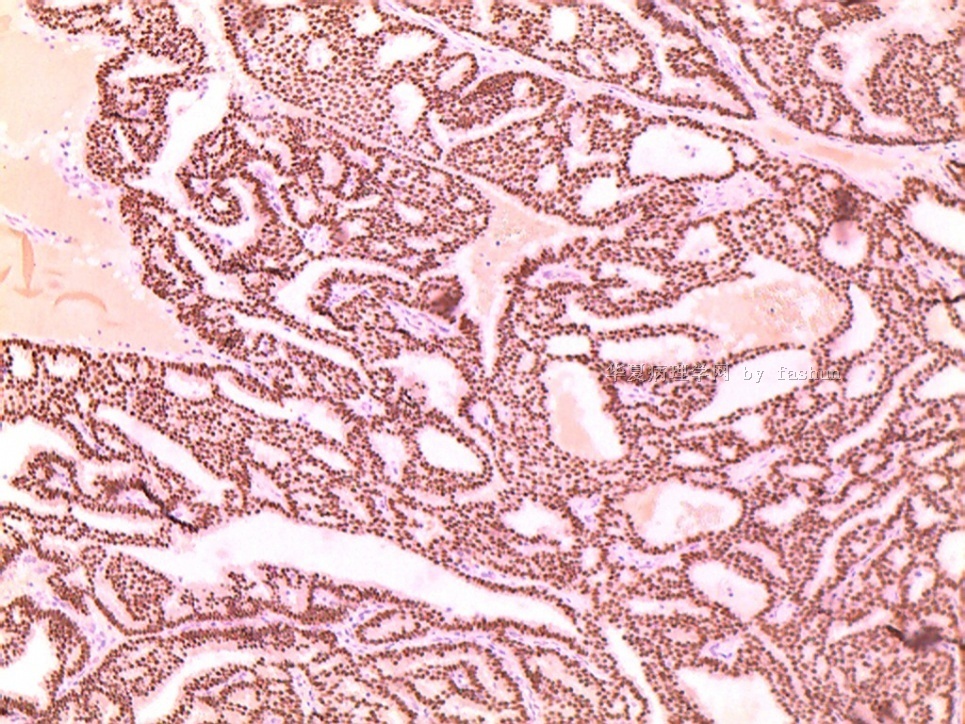
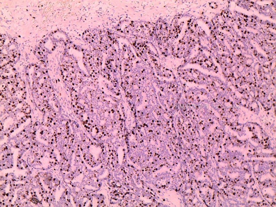
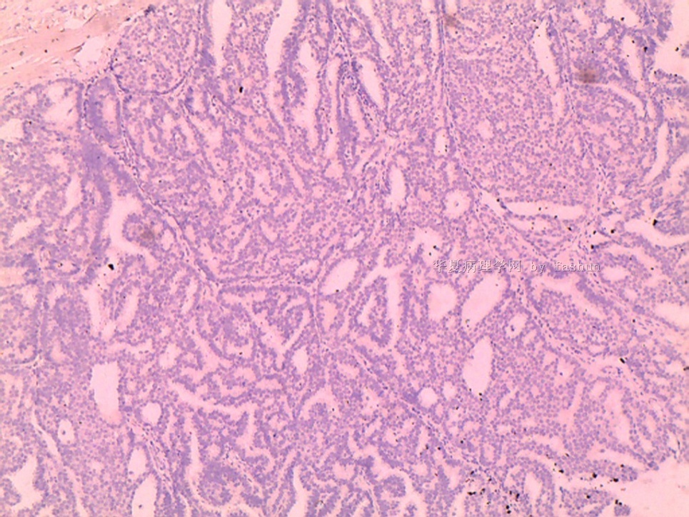
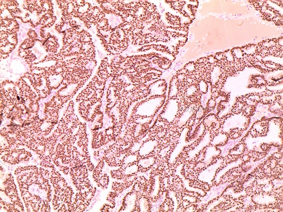
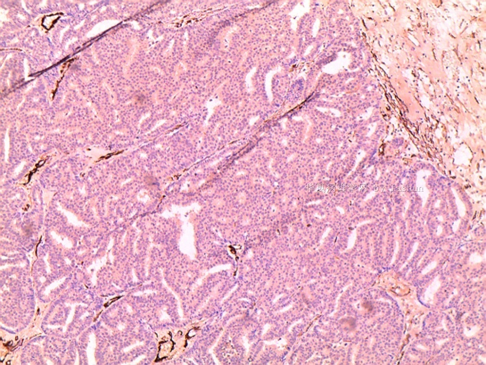
It is an encapsulated papillary ca. Myoepithelial cells absent both within and surrounding the papillary nodules.
Need to read H&E and myoepithelial cell stained slides very carefully to see if foci of unequivocal invasion carcinoma are present.
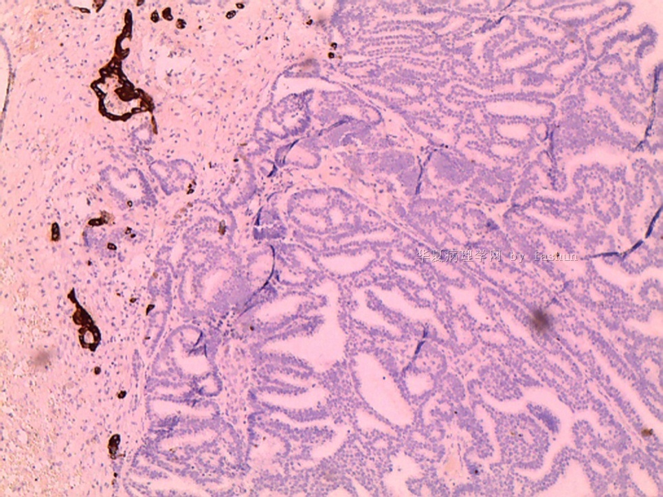
第四张为ck5/6: three small glands without myoepithelial cell surrounding are in the out side of the large encapsulated papillary ca, but the diagnosis of focal invasive ca cannot be made in this photo. It was caused by sectioning.
Good case. Thnak for sharing.
