| 图片: | |
|---|---|
| 名称: | |
| 描述: | |
- 谈东风病例4 Case T0004:30岁女性,阑尾切除标本,是肿瘤吗?
| 姓 名: | ××× | 性别: | female | 年龄: | 30 |
| 标本名称: | |||||
| 简要病史: | abdomen pain | ||||
| 肉眼检查: | a dilated appendiceal mass, 8.5x4.4x3.2cm, cystic appearance with yellow, cloudy mucinous fluid. | ||||
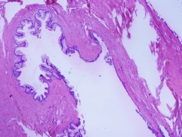
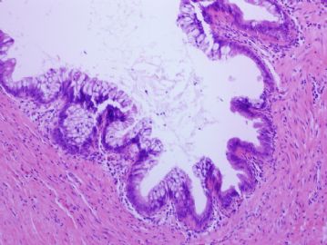
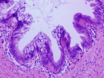
Microscopically, cystic spaces/lesions involves the deep portion of the appendiceal wall, some of which are close to the serosal surface.
Images 1-3: from the mucosal/luminal surface.
Images 4-6: from two cystic lesions close to the serosa.
Impression: a large mucinous mass of the appendix. Microscopically, focal stratification is present, without significant nuclear atypia. Though the lesion involves the subserosal areas (the diverticuli-like changes are frequently in this tumor) , no destructive invasion is identified.
Diagnosis:
Mucinous adenoma with low-grade dysplasia or low-grade mucinous neoplasm: 阑尾低级别黏液性腺瘤
You are doing wonderfully. If this case was perforated, it would be called as "borderline neoplasm with uncertain malignant potential", since it will cause uncureable recurrence later.
-
本帖最后由 于 2010-06-02 23:31:00 编辑


