| 图片: | |
|---|---|
| 名称: | |
| 描述: | |
- 谈东风病例1 Case T0001: 胃肿物
| 姓 名: | ××× | 性别: | female | 年龄: | 72 |
| 标本名称: | mass of the stomach | ||||
| 简要病史: | recent 4 kg weight loss. No dysphagia or GI bleeding | ||||
| 肉眼检查: | A 10x9.0x1.2cm mass at the proximal stomach (3cm below the GE junction). Elevated surface with beef red appearance. No ulcer. Serial sections show the mass is confined within the mucosal layer. | ||||
女性患者,72岁。最近体重减轻4公斤,没有吞咽困难和胃肠道出血。
肉眼检查,距离胃食管交界3cm处、胃近端可见10*9*1.2cm的肿块,表面隆起,呈牛肉红色,无溃疡形成,连续切片显示肿块位于粘膜层。
I've tried to upload the photos several times. Finally 小荷 helps me put them in.
Please be patience with me. I'm still learning and get there.
Many thanks!
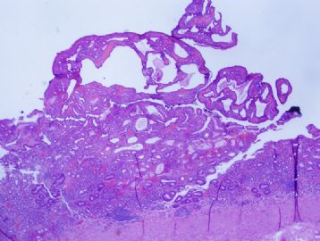
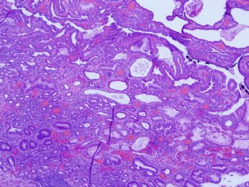
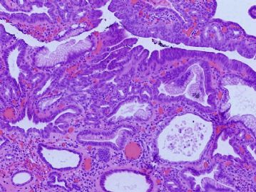
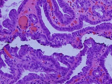
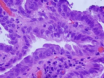
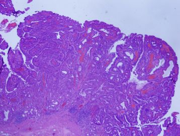
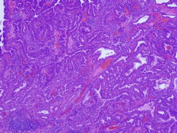
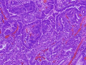
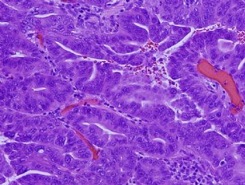
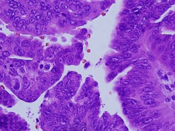
Main clinicopathological features: Elder female with a large mass confined within the mucosal layer. Microscopically, the mass arises in a background of chronic atrophic gastritis with intestinal metaplasia (at the bottom portion of Figure 1 and Figure 2, below). The mass consists of closely packed pyloric gland-type tubules or glands lined by a single layer of columnar or cuboidal epithelium. The cytoplasm is eosinophilic or oncocytic, leading to a "ground-glass" appearance. Intracellular mucin droplets are not identifiable. Low-grade dysplasia is readily seen, and foci of high-grade dysplasia(complex glandular architecture with marked cytological atypia, without desmoplastic stroma) is also seen. Some of the high-grade dysplasia shown in the photos may be called as “intramucosal adenocarcinoma” using Japanese criteria.
Diagnosis:
Pyloric gland adenoma with foci of high-grade dysplasia, without definite invasive adenocarcinoma.
Differential Diagnosis:
The differential diagnosis of this case includes hyperplastic polyp, Menetrier disease, and other gastric adenomas, namely gastric foveolar type adenoma and intestinal type adenoma.
There are overlapping features in pyloric gland adenoma and gastric hyperplastic polyp(GHP). For example, GHP also usually arises in a background of atrophic gastritis. Characteristically, GHP consists of marked elongation, infolding, and branching of the gastric pits leading to a serrated appearance. Mucin-secreting foveolar cells, some resembling goblet cells, line exaggerated, elongated, and distorted pits.In contrast, pyloric gland adenoma characteristically reveals a cytoplasmic "ground-glass" appearance, rather than foveolar-type cells with prominent apical cytoplasmic mucin droplets.
In Menetrier disease, the proliferative epithelium also forms hyperplastic gastric folds, while chronic atrophic gastritis and intestinal metaplasia are absent.
It is essential to distinguish foveolar-type gastric adenoma and pyloric gland adenoma because the latter likely has foci of high grade dysplasia and/or invasive adenocarcinoma. In foveolar type gastric adenoma, the epithelial cells resembling foveolar epithelium similar to that of GHP. Moreover, These lesions usually arise in gastric mucosa which does not show evidence of atrophy or intestinal metaplasia.
Like its counterpart in the coloretum, intestinal-type gastric adenoma usually contain scattered goblet or Paneth cells, though it also often arise in association with chronic atrophic gastritis.
Key Points
**Pyloric gland adenoma is relatively rare gastric epithelial lesion, which is probably under-recognized by pathologists. It can also occurs in non-gastric organs (it has been reported in the gallbladder
and pancreas).
**Pyloric adenoma is usually large ( about 2.0cm and larger) at the time of diagnosis.
**Elder female is the usual patient population.
**Usually it arises in a background of chronic atrophic gastritis with intestinal metaplasia.
**High-grade dysplasia is common, and carefully look for invasive adenocarcinoma is warranted, since reported adenocarcinoma(up to 30% of patients) is usually quite small.
References
Abraham SC, Montgomery EA, Singh VK, et al. Gastric adenomas. Intestinal-type and gastric-type adenomas differ in the risk of adenocarcinoma and presence of background mucosal pathology. Am J Surg Pathol 2002;26:1276-1285.
Vieth M, Kushima R, Borchard F, Stolte M. Pyloric gland adenoma: a clinico-pathological analysis of 90 cases. Virchows Arch 2003;442: 317-321.
Chen ZM, Scudiere JR, Abraham SC, Montgomery E. Pyloric gland adenoma. An entity distinct from gastric foveolar type adenoma. Am J Surg Pathol, 2008,
Vieth M, Kushima R, de Jonge JP, et al. Adenoma with gastric differentiation (so-called pyloric gland adenoma) in a heterotopic gastric corpus mucosa in the rectum. Virchows Arch 2005;446:542-545.
Happy Labor Day (May 1)!
-
本帖最后由 于 2011-04-11 23:07:00 编辑
| 以下是引用xljin8在2010-4-28 21:07:00的发言: |
Excellent differential diagnosis.
I would like to hear more from all of you.
The answer will be posted on April 30.
Thanks.

