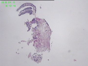
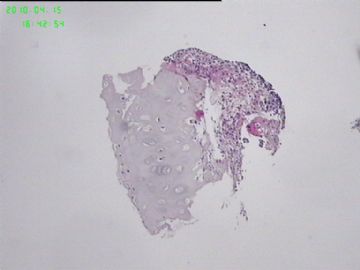
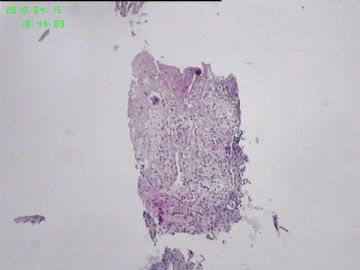
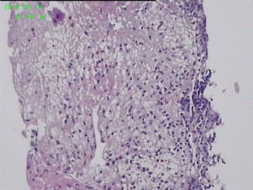
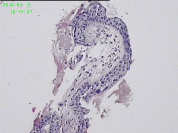
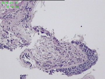
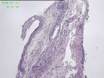
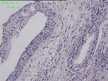
| 图片: | |
|---|---|
| 名称: | |
| 描述: | |
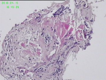
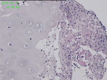
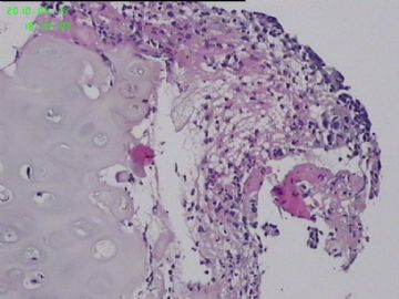
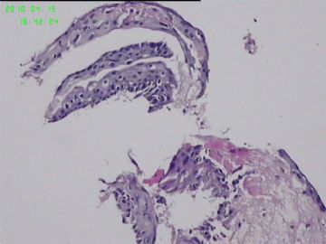
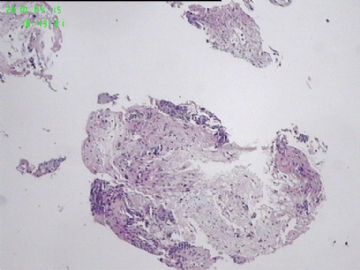
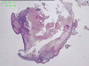
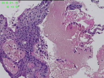
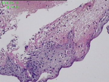
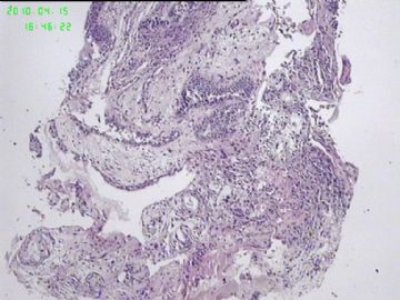
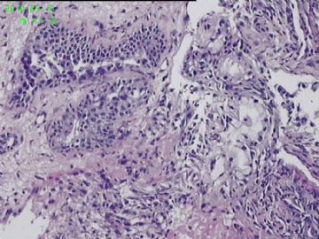
- 右上叶肉芽样新生物,怎么诊断?
请看肺机胎瘤的报道1
1.
Ann Thorac Cardiovasc Surg. 2009 Aug;15(4):247-9.
Primary pulmonary teratoma: Report of a case and the proposition of "bronchotrichosis" as a new term.
Turna A, Ozgül A, Kahraman S, Gürses A, Fener N, Yilmaz V.
Department of Thoracic Surgery, Yedikule Teaching Hospital for Chest Diseases and Thoracic Surgery, Instanbul, Turkeyl.
Abstract
Primary pulmonary teratoma is a very rare disease. Most follow a benign course and are incidental findings during routine chest X-rays. Hair found in sputum or in bronchus detected during bronchoscopy is also a rare condition and is usually caused by mediastinal teratoma. This case report is of a 36-year-old man who presented with halitosis. A fiber-optic bronchoscopy revealed coarse hair originated from the right upper lobe. The patient was successfully treated by right upper lobectomy, and pathology confirmed primary pulmonary teratoma. We recommend that "bronchotrichosis" could be used as a new term for such a sign.

- 王军臣
请见肺畸胎瘤报道2
J Bras Pneumol. 2007 Oct;33(5):612-5.
Intrapulmonary teratoma.
[Article in English, Portuguese]
Faria RA, Bizon JA, Saad Junior R, Dorgan Neto V, Botter M, Saieg MA.
Faculdade de Ciências Médicas da Santa Casa de São Paulo - FCMSCSP, Santa Casa School ofMedical Sciences in São Paulo - São Paulo (SP) Brazil. rickpreto@zipmail.com.br
Abstract
Case report of a 49-year-old man, presenting chest pain and bloody sputum for six months. Chest X-ray and computed tomography scan showed opacification on the left upper lobe. The bronchoscopy showed bronchial hemorrhage in the lingular bronchial segment. Due to diagnostic and therapeutic needs, this patient underwent a left inframammilary thoracotomy. The anatomopathological analysis of the surgical sample revealed an intrapulmonary teratoma. The patient presented favorable evolution and is now under outpatient follow-up treatment.

- 王军臣
请见肺畸胎瘤报道3
Ann Thorac Surg. 2007 Mar;83(3):1194-6.
Intrapulmonary teratoma: an exceptional disease.
Rana SS, Swami N, Mehta S, Singh J, Biswal S.
Department of Cardio-thoracic & Vascular Surgery, Post Graduate Institute of Medical Education & Research, Chandigarh, India.
Abstract
Intrathoracic teratomas almost always occur in the mediastinum, but occasionally, they may be found in the lung as intrapulmonary teratomas. Intrapulmonary teratomas have histologic findings that are similar to those of teratoma from other sites. Two successive patients with intrapulmonary teratomas presented to us in a variable manner. The clinical and radiologic features and the histopathologic findings are presented, and the relevant literature is discussed.

- 王军臣

