| 图片: | |
|---|---|
| 名称: | |
| 描述: | |
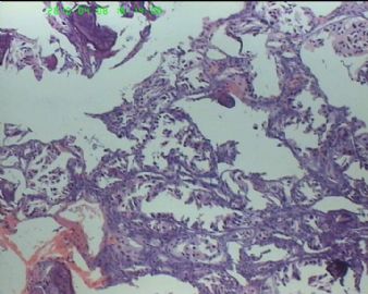
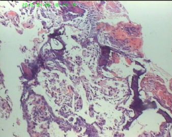
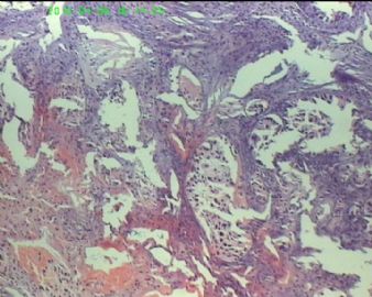
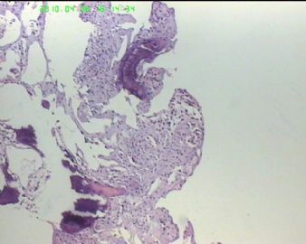
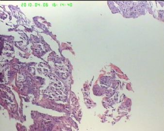
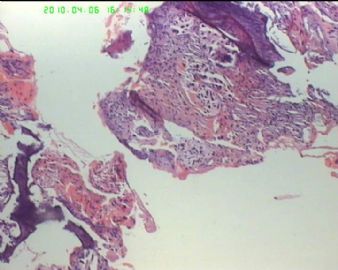
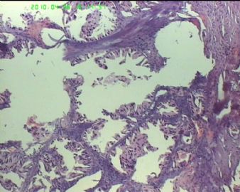
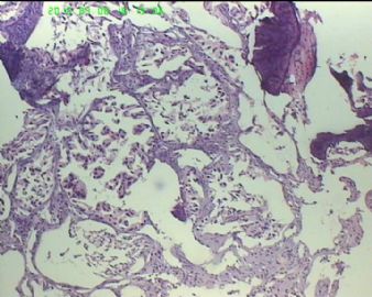
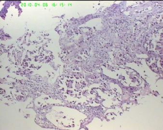
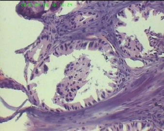
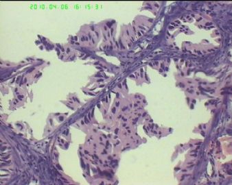
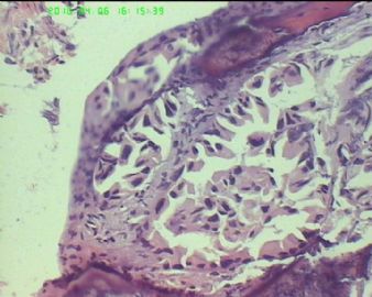
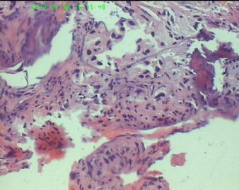
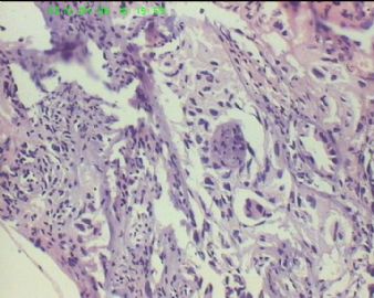
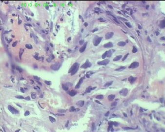
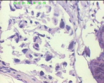
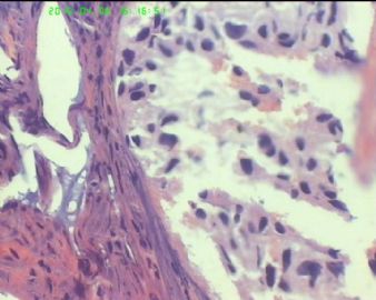
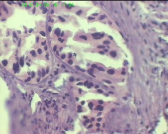
- 55岁 蝶窦内斜坡硬膜外质硬肿物伴骨破坏,供血丰富
| 以下是引用mjma在2010-4-8 1:13:00的发言: Most of the tissue resected appears cauterized, probably due to rich vascularity and intraoperative bleeding. I see epithelial structures lined by somewhat atypical glandular epithelia. In the sphenoid sinus and clivus region, these would certainly suggest an adenocarcinoma of metastatic origin. However, examination of less cauterized areas, if any, is essential. Immunohistochemistry (AE1) may help. The photos do not look like that from a meningioma or pituitary adenoma. |
-
zhoubingjuan 离线
- 帖子:261
- 粉蓝豆:366
- 经验:590
- 注册时间:2007-05-30
- 加关注 | 发消息
-
Most of the tissue resected appears cauterized, probably due to rich vascularity and intraoperative bleeding. I see epithelial structures lined by somewhat atypical glandular epithelia. In the sphenoid sinus and clivus region, these would certainly suggest an adenocarcinoma of metastatic origin. However, examination of less cauterized areas, if any, is essential. Immunohistochemistry (AE1) may help. The photos do not look like that from a meningioma or pituitary adenoma.

聞道有先後,術業有專攻

