


| 图片: | |
|---|---|
| 名称: | |
| 描述: | |
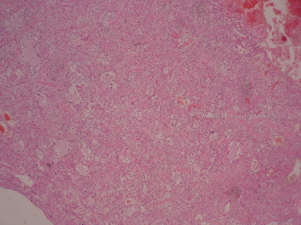
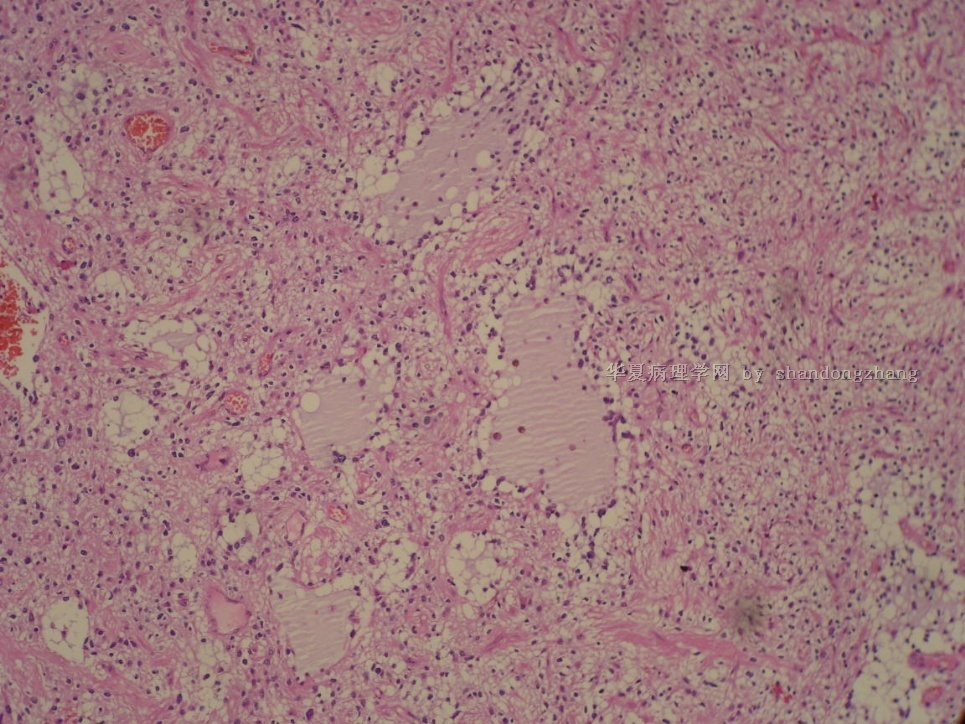
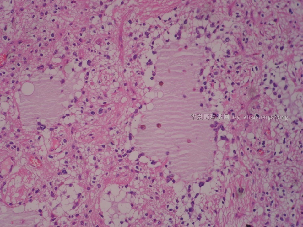
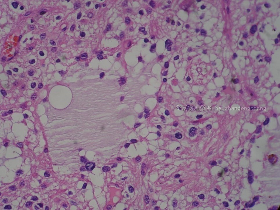
- 20100331-右额叶占位
-
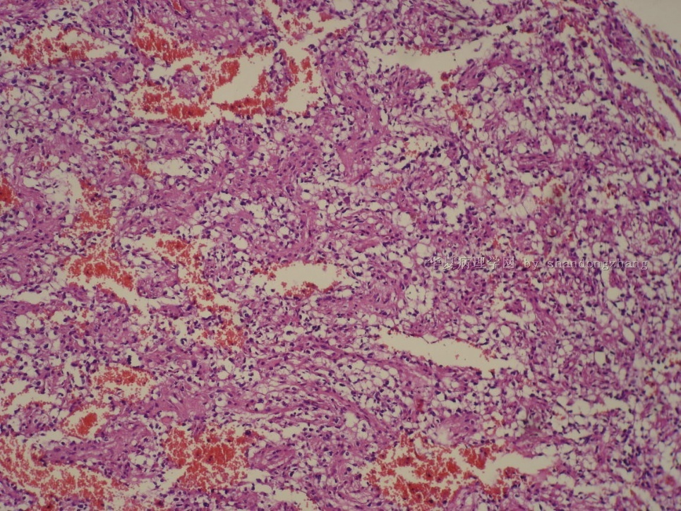
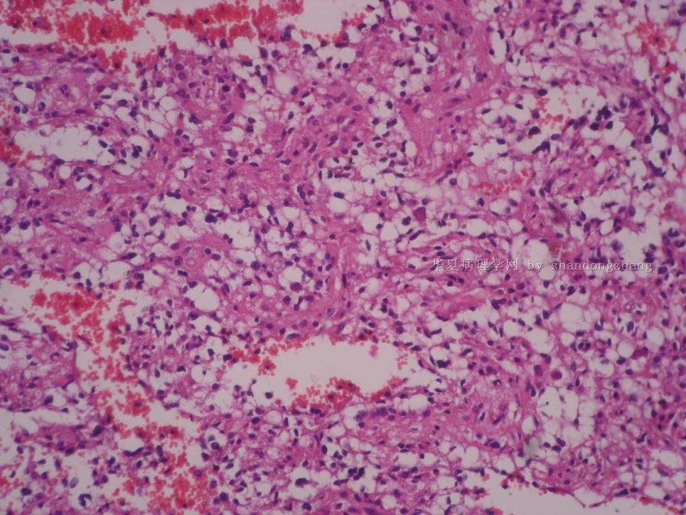
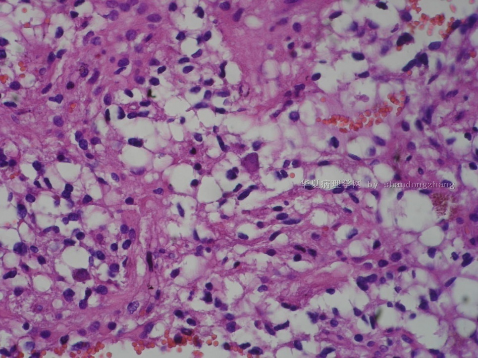
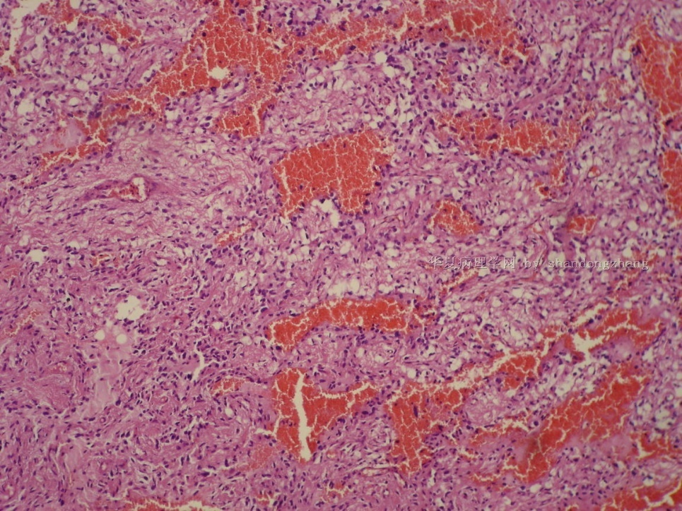
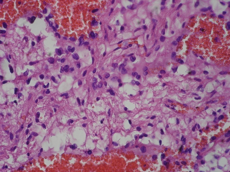
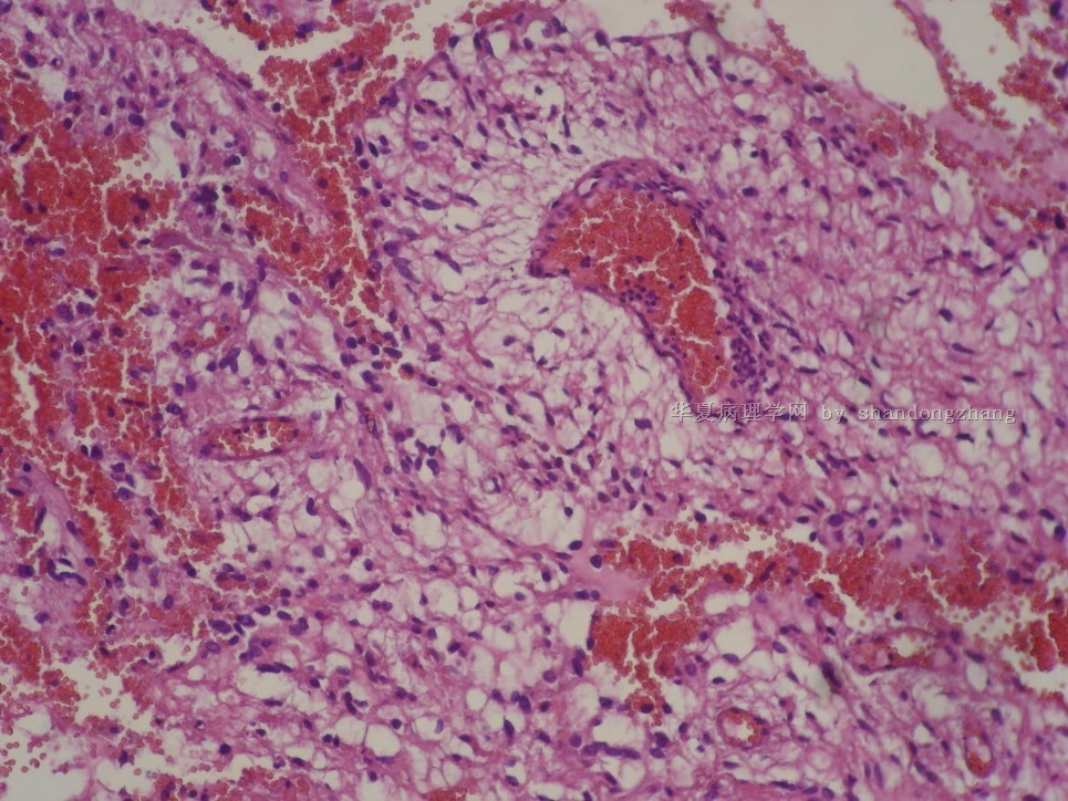
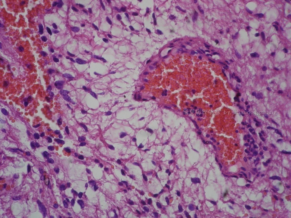
This is an interesting case. The neoplastic cells appear glial, and many have oval and relatively uniform nuclei surrounded by perinuclear halos. Some neoplastic cells are more elongated and astrocytic. There is a distinctive biphasic growth pattern - eosinophilic fibrillary areas alternating with mucoinous microcysts containing loosely arranged cells with perinuclear halos. There are scattered calcospherites strongly suggestive of a low grade neoplasm. Occasional isolated giant cells with large and hyperchromatic nuclei are present focally. In some areas neoplastic cells are found between a network of small vessels suggestive of that seen in oligodendroglioma. I do not find mitotic figures, so presumably they are very rare. I do not see necrosis. vascular/endothelial proliferation or features of cytologic anaplasia. My first impression is that this is WHO grade I pilocytic astrocytoma with hemorrhage. I would look for the following features to confirm or support my impression - eosinophilic granular bodies and Rosenthal fibers. If neither structures are found, I would carefully search and count mitotic figures. If they are fewer than 1~2 per 10 high-power fields, this is still likely a pilocytic astrocytoma. Even if mitotic figures are more than 2 per 10 high-power fields, it is still a pilocytic astrocytoma (perhaps a rare case of atypical pilocytic astrocytoma) if either eosinophilic granular bodies or Rosenthal fibers are present. I do ot think this is oligodendroglioma or fibrillary astrocytoma.

聞道有先後,術業有專攻
-
从颅内原发性肿瘤来看,以透明细胞为主的肿瘤主要有少突胶质细胞瘤,DNT,PA(毛细胞星形细胞瘤),透明细胞性脑膜瘤,透明细胞性室管膜瘤,血管母细胞瘤等。就此例形态来看,间质血管不支持少突胶质细胞瘤,透明细胞性脑膜瘤/室管膜瘤未见相应的特点,DNT不太象(未见特异性胶质神经元成份),血管母细胞瘤形态上不支持,剩下来主要是考虑PA,PA可以局部或全部表现似少突胶质细胞瘤,但仍保留PA的主要特点,如双相结构、EGB及Rosenthal fibers等。从病史上看,甲状腺及子宫有肿物切除史,形态上主要与甲状腺透明细胞癌及子宫的PEComa鉴别,但均不太支持,IHC抗体选择时可考虑用TG、TTF1及HMB45。
-
zhoubingjuan 离线

- 帖子:261
- 粉蓝豆:366
- 经验:590
- 注册时间:2007-05-30
- 加关注 | 发消息
| 以下是引用mjma在2010-3-31 11:15:00的发言: This is an interesting case. The neoplastic cells appear glial, and many have oval and relatively uniform nuclei surrounded by perinuclear halos. Some neoplastic cells are more elongated and astrocytic. There is a distinctive biphasic growth pattern - eosinophilic fibrillary areas alternating with mucoinous microcysts containing loosely arranged cells with perinuclear halos. There are scattered calcospherites strongly suggestive of a low grade neoplasm. Occasional isolated giant cells with large and hyperchromatic nuclei are present focally. In some areas neoplastic cells are found between a network of small vessels suggestive of that seen in oligodendroglioma. I do not find mitotic figures, so presumably they are very rare. I do not see necrosis. vascular/endothelial proliferation or features of cytologic anaplasia. My first impression is that this is WHO grade I pilocytic astrocytoma with hemorrhage. I would look for the following features to confirm or support my impression - eosinophilic granular bodies and Rosenthal fibers. If neither structures are found, I would carefully search and count mitotic figures. If they are fewer than 1~2 per 10 high-power fields, this is still likely a pilocytic astrocytoma. Even if mitotic figures are more than 2 per 10 high-power fields, it is still a pilocytic astrocytoma (perhaps a rare case of atypical pilocytic astrocytoma) if either eosinophilic granular bodies or Rosenthal fibers are present. I do ot think this is oligodendroglioma or fibrillary astrocytoma. |


