| 图片: | |
|---|---|
| 名称: | |
| 描述: | |
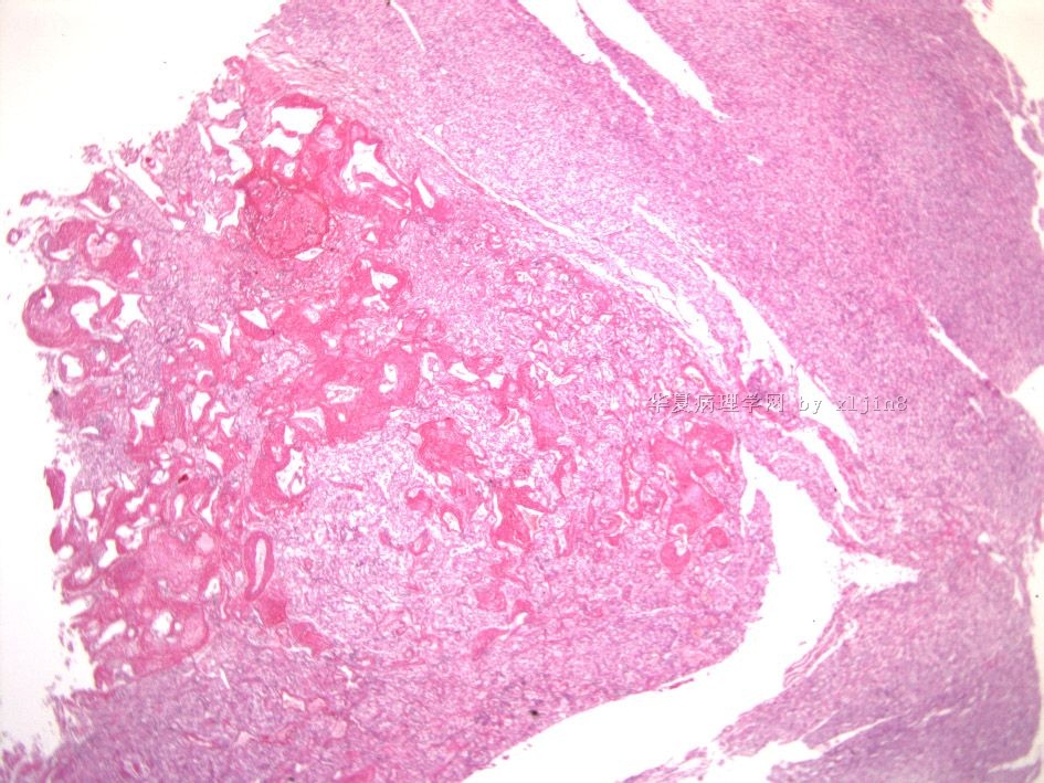
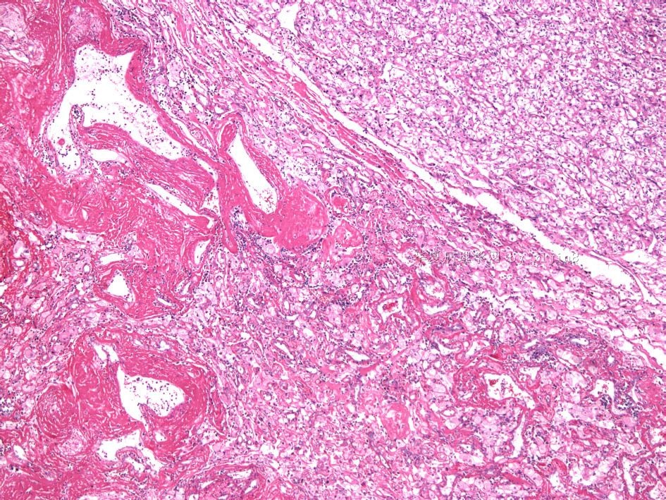
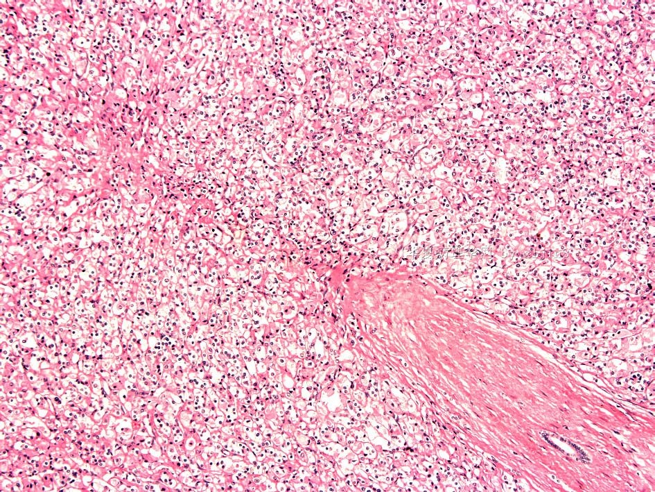
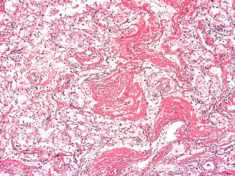
- 男/35岁 黑色素性Xp11易位肾癌?(上海市疑难病例读片会2010#1长海医院病例)
| 姓 名: | ××× | 性别: | 年龄: | ||
| 标本名称: | |||||
| 简要病史: | 体检发现右肾肿瘤一周 | ||||
| 肉眼检查: | 肾脏一个,9.5x5x3cm,切面中上级见一肿块,3x3x2.5cm, 灰白灰黄色,部分暗红色,边界较清楚。位于肾实质内,累及肾包膜。 | ||||
图片9 IHC 标记- HMB-45;
图片10-11 肿瘤与正常肾脏 CK7;
图12 肿瘤周围正常肾脏 CK7;
名称:图1
描述:图1
名称:图2
描述:图2
名称:图3
描述:图3
名称:图4
描述:图4
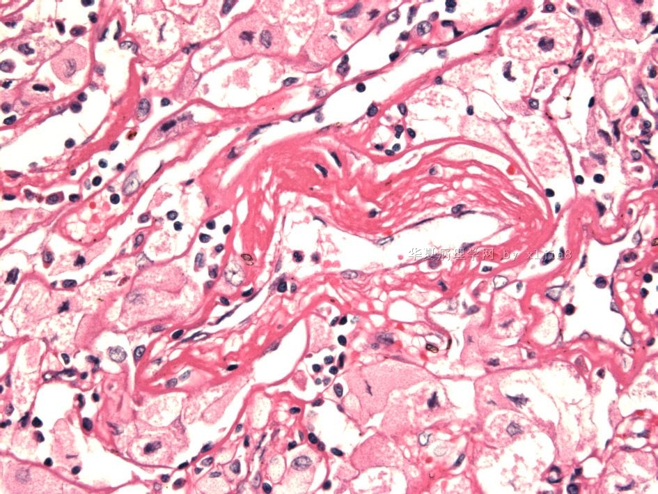
名称:图5
描述:图5
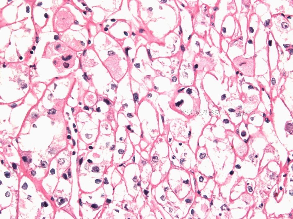
名称:图6
描述:图6
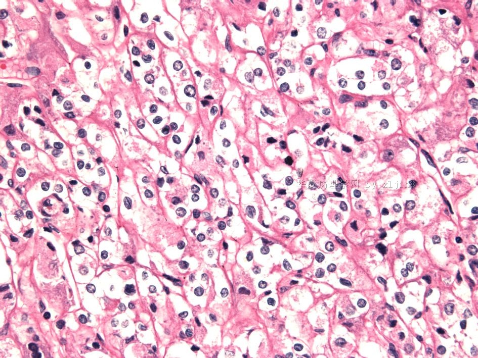
名称:图7
描述:图7
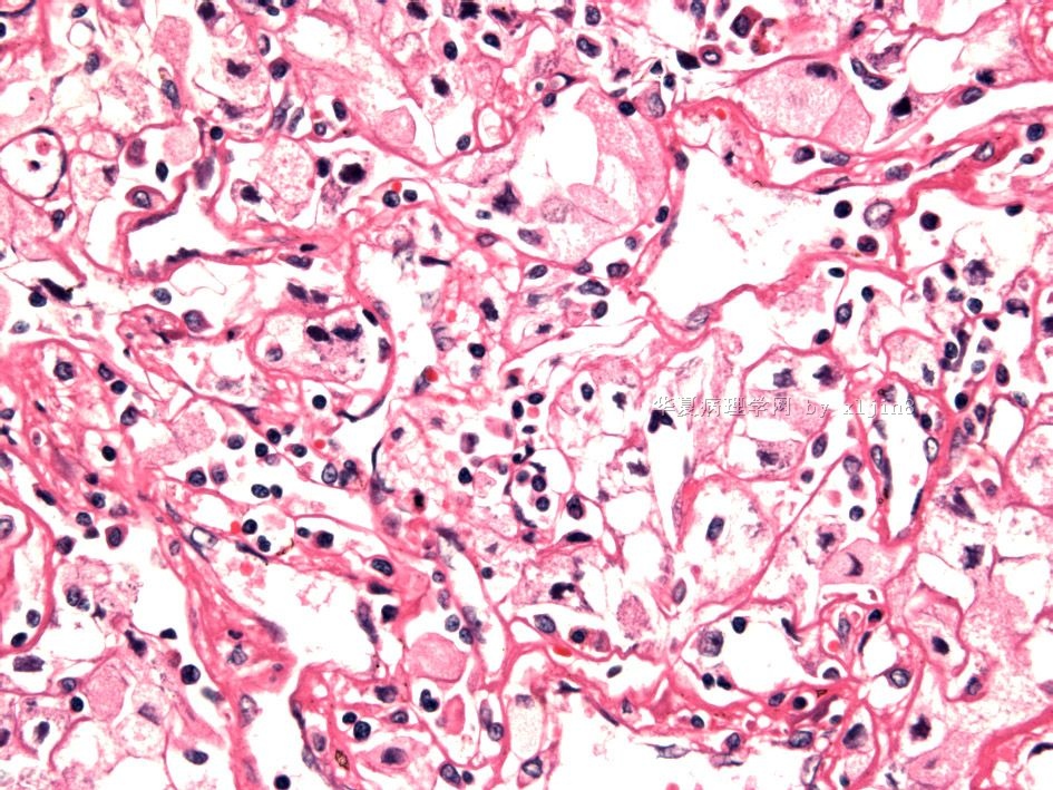
名称:图8
描述:图8
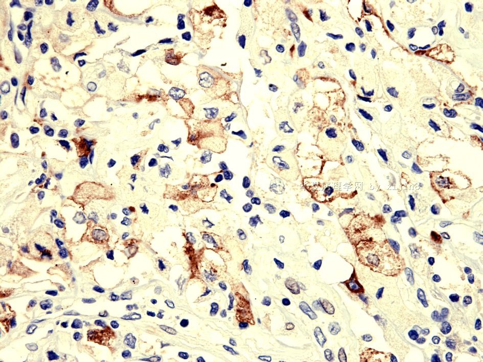
名称:图9
描述:图9
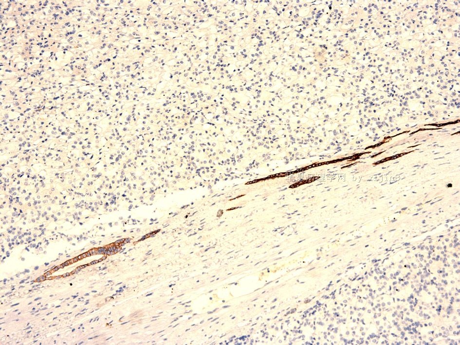
名称:图10
描述:图10
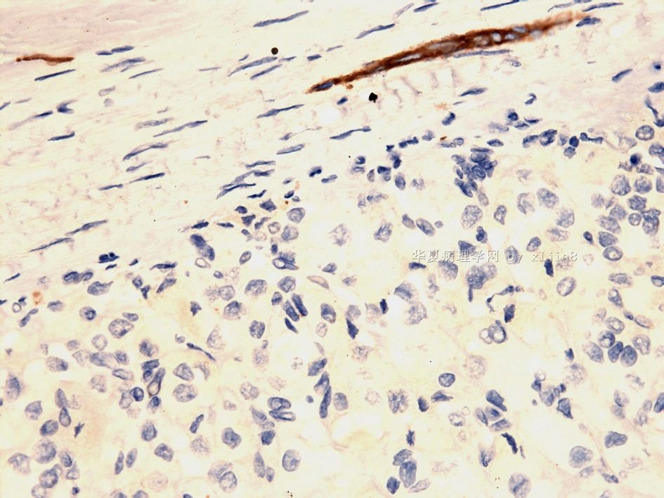
名称:图11
描述:图11
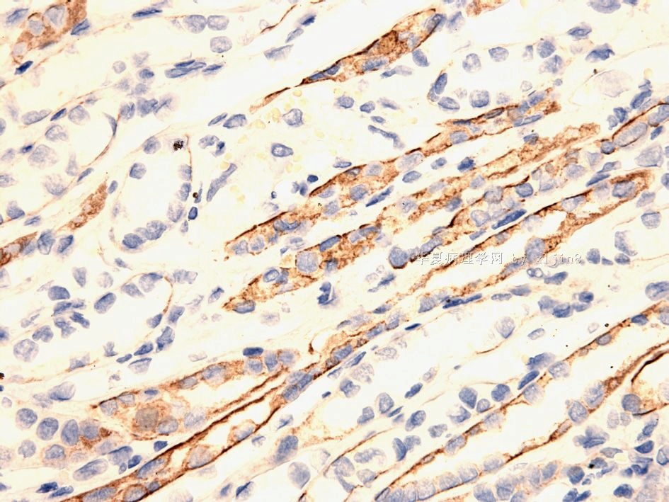
名称:图12
描述:图12
-
本帖最后由 于 2010-04-01 02:56:00 编辑

- xljin8
-
本帖最后由 XLJin8 于 2011-09-07 04:46:43 编辑
如果要归纳黑色素性Xp11易位肾癌的临床病理特征,大致如下:
1)青少年患者,一般小于25岁;
2)HE形态更像肾细胞癌;
3)血管周围有明显的纤维素样沉积,
4)无发育不良的厚壁血管、无脂肪细胞;
5)一般肾细胞癌标记阴性;
6)平滑肌和脂肪细胞标记阴性;
7)HMB-45阳性;

- xljin8
-
本帖最后由 XLJin8 于 2011-09-01 21:10:50 编辑
| 以下是引用xljin8在2010-4-1 2:44:00的发言:
1. Chang IW, Huang HY, Sung MT. Melanotic Xp11 translocation renal cancer: a case with PSF-TFE3 gene fusion and up-regulation of melanogenetic transcripts. Am J Surg Pathol. 2009;33:1894-901. Department of Pathology, Chang Gung Memorial Hospital-Kaohsiung Medical Center, 2. Argani P, Aulmann S, Karanjawala Z, Fraser RB, Ladanyi M, Rodriguez MM. Department of Pathology, The Johns Hopkins Hospital , The Johns Hopkins University, Baltimore, MD 21231-2410, USA. pargani@jhmi.edu We describe 2 cases of malignant melanotic epithelioid renal neoplasms bearing |
By immunohistochemistry, the neoplastic cells labeled for melanocytic markers HMB45 and Melan A, but not for S100 protein, MiTF, or any epithelial marker (cytokeratins, epithelial membrane antigen), renal tubular marker (CD10, PAX8,PAX2, RCC Marker) or muscle marker (actin, desmin).
WHO分类是2004年版,书中描述的是t(Xp11.2) ,与TFE3形成融合基因。而本病例是2009年才报道,请参考上述文献。

- xljin8
-
本帖最后由 于 2010-04-01 02:58:00 编辑
| 以下是引用xljin8在2010-4-1 2:44:00的发言:
1. Chang
IW, Huang HY, Sung MT. Melanotic Xp11 translocation renal cancer: a case
with PSF-TFE3 gene fusion and up-regulation of melanogenetic transcripts. Am J
Surg Pathol. 2009;33:1894-901. Department
of Pathology, Chang Gung Memorial Hospital-Kaohsiung Medical Center, 2. Argani
P, Aulmann S, Karanjawala Z, Fraser RB, Ladanyi M, Rodriguez MM. Department
of Pathology, The Johns Hopkins Hospital , The Johns Hopkins University,
Baltimore, MD 21231-2410, USA. pargani@jhmi.edu We describe
2 cases of malignant melanotic epithelioid renal neoplasms bearing |

- xljin8


