| 图片: | |
|---|---|
| 名称: | |
| 描述: | |
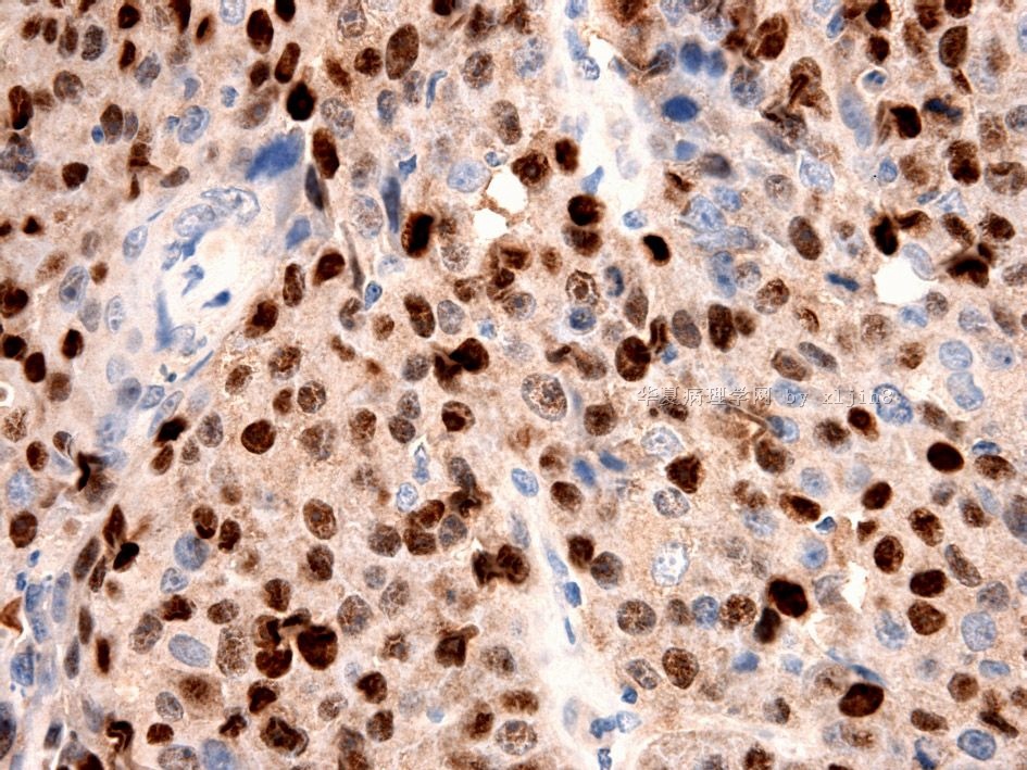
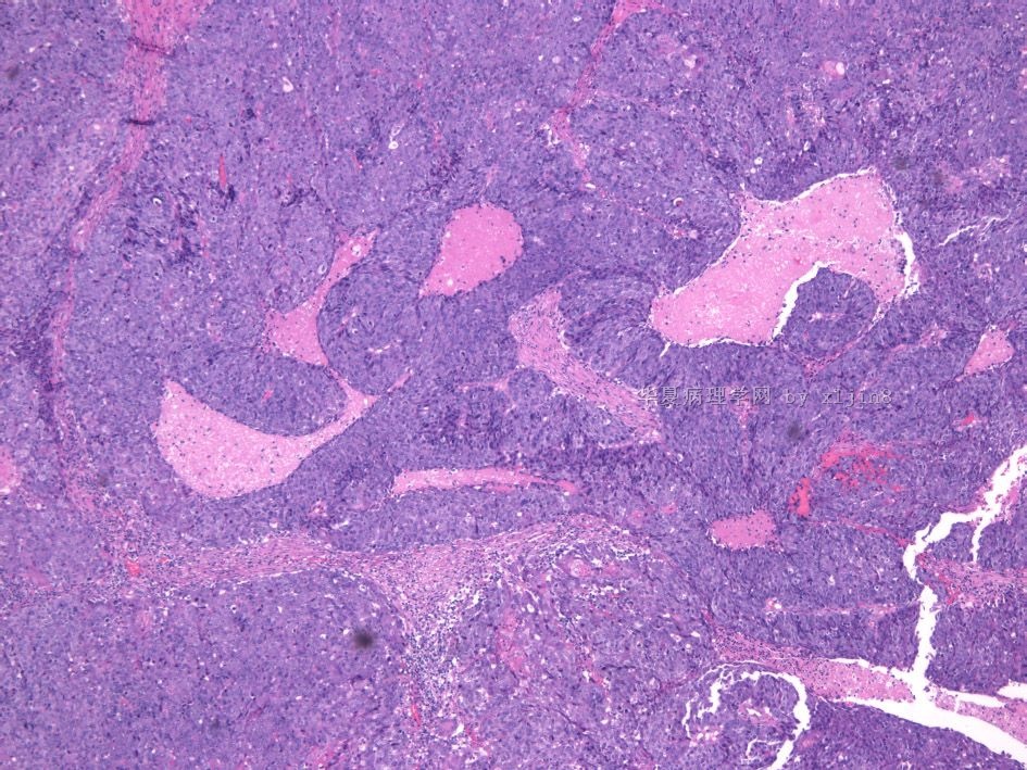
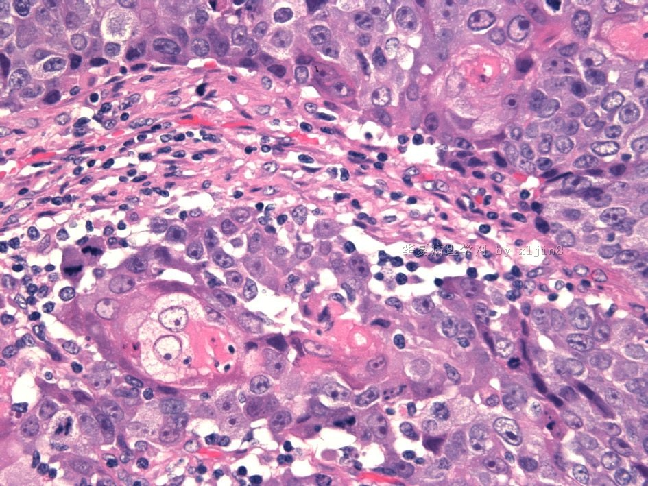
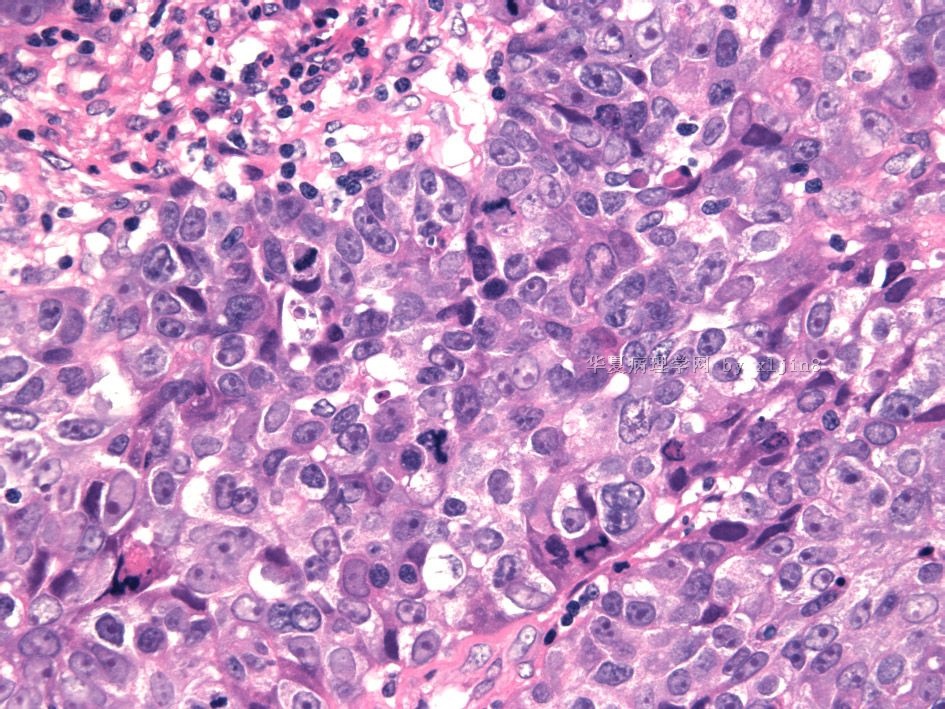
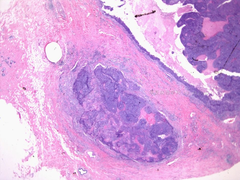
- B2654女/58岁 左乳腺癌 分类?(IHC结果2010-5-3)
| 姓 名: | ××× | 性别: | 年龄: | ||
| 标本名称: | |||||
| 简要病史: | 左乳腺肿块8月余,无痛。最近明显增大。 | ||||
| 肉眼检查: | 乳腺组织5.5X5X4.5 CM, 切面见灰白色肿块,直径2.3cm,位于血性囊腔内。 | ||||
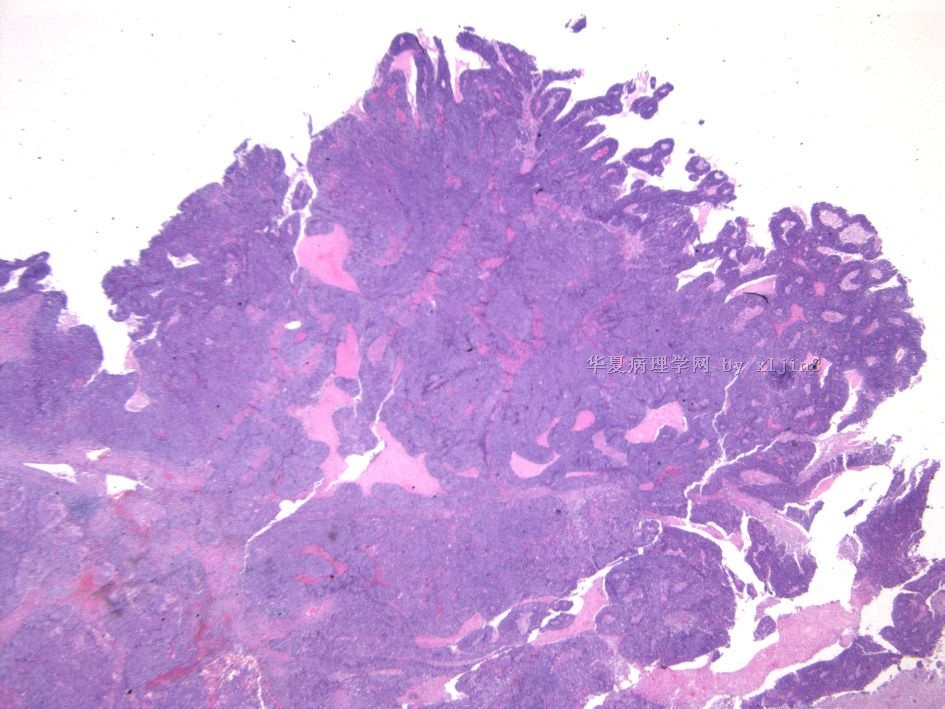
名称:图1
描述:图1
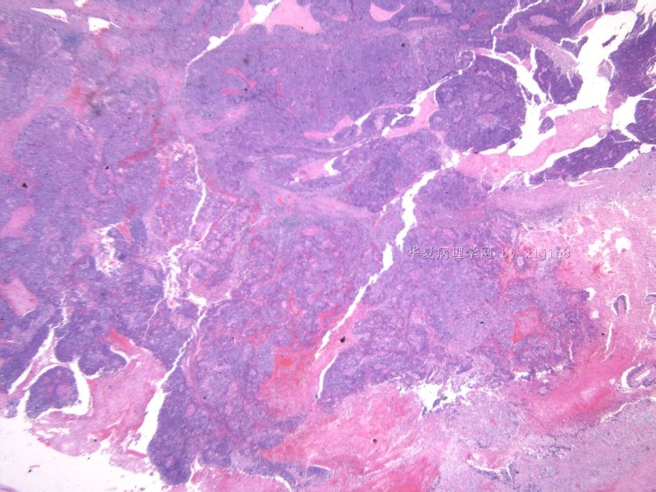
名称:图2
描述:图2
名称:图3
描述:图3
名称:图4
描述:图4
名称:图5
描述:图5
名称:图6
描述:图6
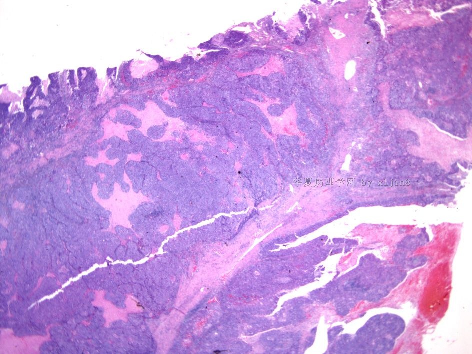
名称:图7
描述:图7
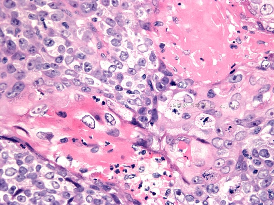
名称:图8
描述:图8
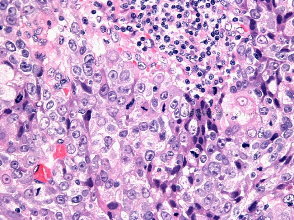
名称:图9
描述:图9
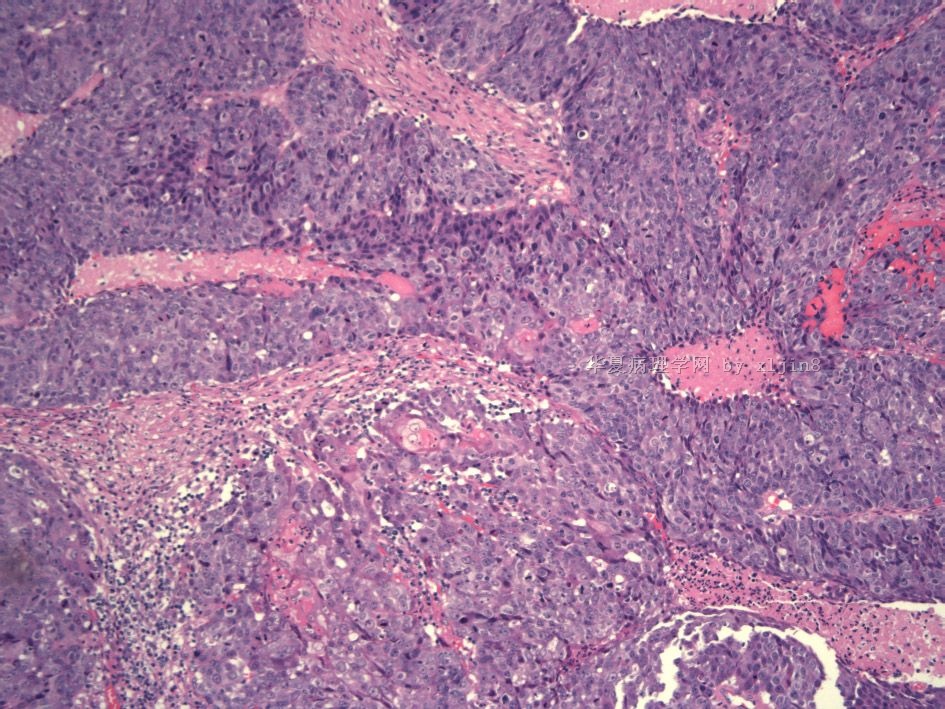
名称:图10
描述:图10
-
本帖最后由 于 2010-05-03 08:34:00 编辑

- xljin8
Do not matter what we call it is a high grade ca. The treatment and prognosis will be the same whatever we call.
If it is my case I will call invasive ductal carcinoma, histologic grading 3 (tubual formation-3, nuclear atypia-3, mitotic activity-3; total score 9/9).
I will not call medullary ca for this case because the infiltrating margins and cytomorphology of the tumor.
太精彩了,Zhao老师您太棒了,我好崇拜您哦!

- 老林
Should stain ER/PR/Her2. If it is triple negative tumor plus some basal marker positive (p63+ already), it is a basal-like carcinoma. Morphology is very classic.
I guess it is invasive ductal ca with basal-like phenotype.
Do not call 髓样癌 for this kind of case.
可能是一种高级别的尿路上皮癌. It can be, but common thing is common.
化生性癌. It is right. Most of 化生性癌are basal-like ca. They are different classication systems.
-
本帖最后由 于 2010-03-29 10:05:00 编辑
1. 乳腺化生性癌是一个总称,涉及一组不同类型的肿瘤,本组肿瘤的特点为腺癌与明显的梭形细胞、鳞状细胞和(或)间叶组织分化区域并存,化生的梭形细胞癌和鳞状细胞癌可以单独存在,不伴有可识别的腺癌成分。根据肿瘤组织学形态可分为多种亚型:
纯上皮化生癌
鳞状细胞癌
腺癌伴梭形细胞化生
腺鳞癌
粘液表皮样癌
上皮/间叶混合性化生性癌
2.大体检查:
质硬,界清,实性;
鳞化或软骨分化时,切面呈珍珠白色或质硬光亮区;
在大的鳞状细胞癌切面可见单个大囊腔或多发小囊腔。
3.鳞状细胞癌的组织病理学:
组织学似良性的高分化细胞被覆在囊腔面,高分化的细胞常形成囊腔。当肿瘤细胞放散出来并浸润周围间质形态变为梭形,失去鳞状细胞特点,明显的间质细胞反应常与梭形细胞鳞癌混合。
4.鳞状细胞癌免疫表型:
CK5,CK34BE12,但不表达血管内皮标志物。几乎所有的鳞状细胞癌均不表达ER/PR.


