共1页/17条首页上一页1下一页尾页
回复:17 阅读:4215
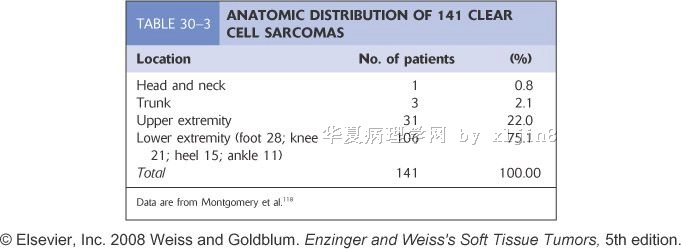
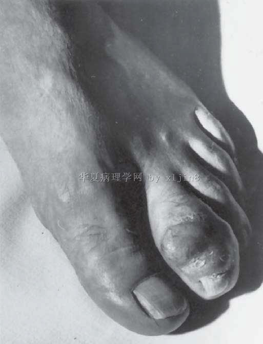
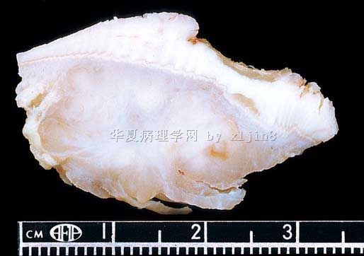
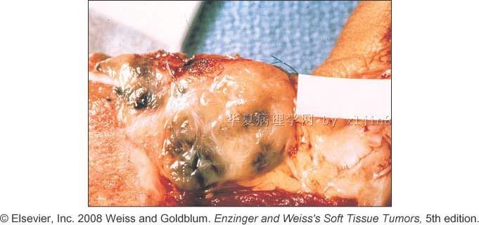
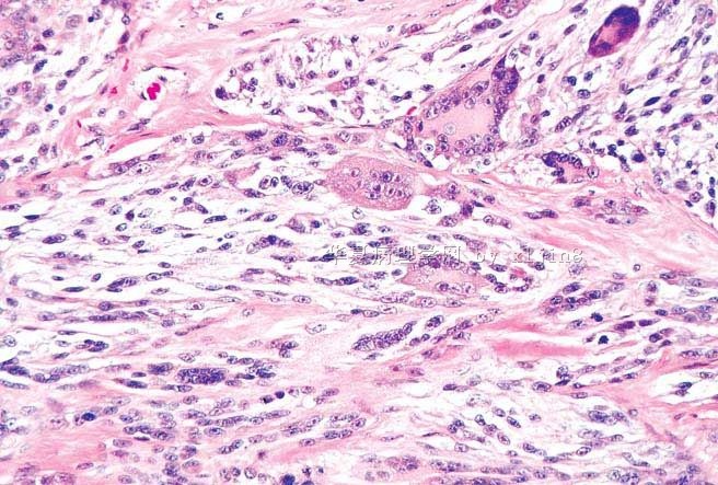
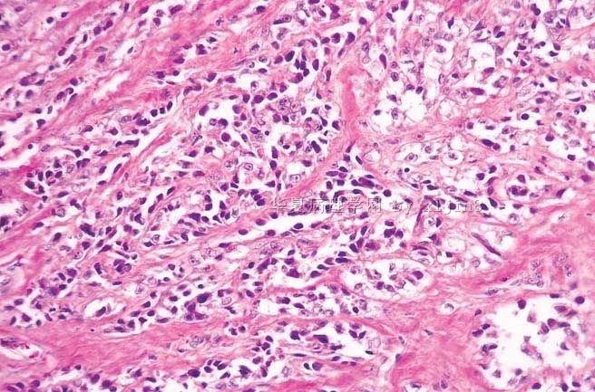
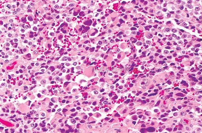
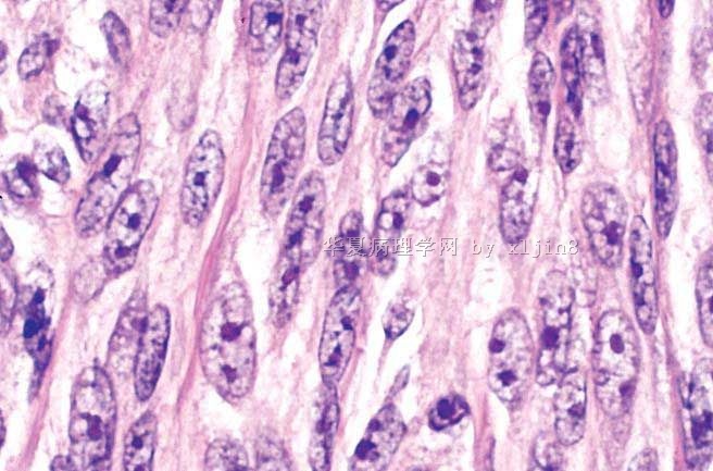
软组织透明细胞肉瘤的鉴别诊断讨论(附Enzinger & Weiss's 软组织肿瘤5版中的CCS图谱)

xljin8 离线

帖子:2275
粉蓝豆:1535
经验:2417
注册时间:2009-03-04
加关注  |  发消息
楼主 发表于 2010-03-25 05:06|举报|关注(2)
浏览排序[ 顺序 逆序 楼主 支持 精彩 ]  快捷回复
姓    名: ××× 性别:   年龄:  
标本名称:  
简要病史:  
肉眼检查:  
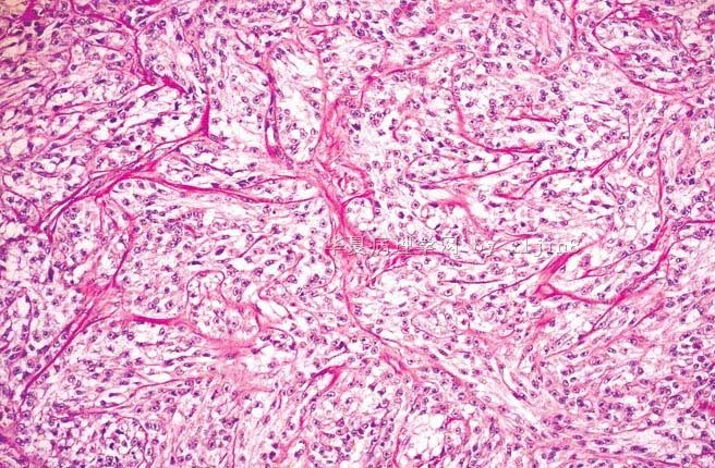
软组织透明细胞肉瘤的鉴别诊断讨论(附Enzinger & Weiss's 软组织肿瘤5版中的CCS图谱)图1
名称:图1
描述:图1
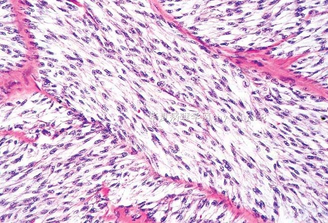
软组织透明细胞肉瘤的鉴别诊断讨论(附Enzinger & Weiss's 软组织肿瘤5版中的CCS图谱)图2
名称:图2
描述:图2
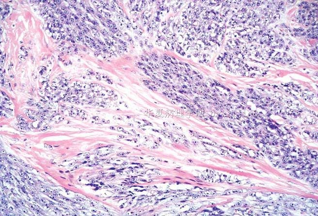
软组织透明细胞肉瘤的鉴别诊断讨论(附Enzinger & Weiss's 软组织肿瘤5版中的CCS图谱)图3
名称:图3
描述:图3
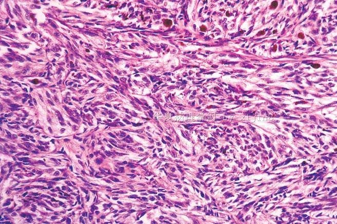
软组织透明细胞肉瘤的鉴别诊断讨论(附Enzinger & Weiss's 软组织肿瘤5版中的CCS图谱)图4
名称:图4
描述:图4
软组织透明细胞肉瘤的鉴别诊断讨论(附Enzinger & Weiss's 软组织肿瘤5版中的CCS图谱)图5
名称:图5
描述:图5
软组织透明细胞肉瘤的鉴别诊断讨论(附Enzinger & Weiss's 软组织肿瘤5版中的CCS图谱)图6
名称:图6
描述:图6
软组织透明细胞肉瘤的鉴别诊断讨论(附Enzinger & Weiss's 软组织肿瘤5版中的CCS图谱)图7
名称:图7
描述:图7
软组织透明细胞肉瘤的鉴别诊断讨论(附Enzinger & Weiss's 软组织肿瘤5版中的CCS图谱)图8
名称:图8
描述:图8
软组织透明细胞肉瘤的鉴别诊断讨论(附Enzinger & Weiss's 软组织肿瘤5版中的CCS图谱)图9
名称:图9
描述:图9
软组织透明细胞肉瘤的鉴别诊断讨论(附Enzinger & Weiss's 软组织肿瘤5版中的CCS图谱)图10
名称:图10
描述:图10
软组织透明细胞肉瘤的鉴别诊断讨论(附Enzinger & Weiss's 软组织肿瘤5版中的CCS图谱)图11
名称:图11
描述:图11
软组织透明细胞肉瘤的鉴别诊断讨论(附Enzinger & Weiss's 软组织肿瘤5版中的CCS图谱)图12
名称:图12
描述:图12
标签:
本帖最后由 于 2010-03-26 05:50:00 编辑
0
signature
xljin8
添加参考诊断
×参考诊断
  

XLJin8 离线

帖子:2275
粉蓝豆:1535
经验:2417
注册时间:2009-03-04
加关注  |  发消息
1 楼    发表于2010-03-25 05:11:00举报|引用
返回顶部 | 快捷回复
最近有二例软组织透明细胞肉瘤,有人提问:如何进行透明细胞肉瘤与恶性黑色素瘤的鉴别诊断?请大家发表意见,谢谢!
0
回复
signature
xljin8

njwbhuang 离线

帖子:712
粉蓝豆:2265
经验:1404
注册时间:2007-11-23
加关注  |  发消息
2 楼    发表于2010-03-25 10:03:00举报|引用
返回顶部 | 快捷回复

 Cutaneous Clear Cell Sarcoma: A Clinicopathologic,Immunohistochemical, and Molecular Analysis of 12 Cases Emphasizing its Distinction from Dermal Melanoma

Markus Hantschke, MD,* Thomas Mentzel, MD,* Arno Ru¨ tten, MD,* Gabriele Palmedo, PhD,* Eduardo Calonje, MD,w Alexander J. Lazar, MD,z and Heinz Kutzner, MD*

(Am J Surg Pathol 2010;34:216–222)

该文中作者讨论中提到免疫组化在二者的鉴别诊断中没有价值,但有一些组织形态学可以区分:透明细胞肉瘤在整个肿瘤中索性细胞呈一致的束状排列,其间由纤细的纤维分隔,间质大多数透明硬化和网状。而黑色素瘤病变内不止一种细胞形态。多核巨细胞在这2种病变中均可存在,但在CCS中较散在,核位于周边呈花环状,形态较一致,而黑色素瘤中多核巨细胞分布无规律,且核多形性。如有条件,进行EWS-ATF1和EWS-CREB1检测非常有价值。

0
回复

XLJin8 离线

帖子:2275
粉蓝豆:1535
经验:2417
注册时间:2009-03-04
加关注  |  发消息
3 楼    发表于2010-03-25 18:36:00举报|引用
返回顶部 | 快捷回复

非常感谢Dr.Huang 提供的最新文献。

请教一个问题: "Cutaneous clear cell sarcoma" 与"Clear cell sarcoma of tendon and aponeurosis" 是否是指同一种肿瘤?
0
回复
signature
xljin8

njwbhuang 离线

帖子:712
粉蓝豆:2265
经验:1404
注册时间:2007-11-23
加关注  |  发消息
4 楼    发表于2010-03-25 20:56:00举报|引用
返回顶部 | 快捷回复

 金主任,根据文献,它们是同一种肿瘤。

0
回复

天山望月 离线

帖子:4902
粉蓝豆:83
经验:5092
注册时间:2007-02-03
加关注  |  发消息
5 楼    发表于2010-03-25 21:59:00举报|引用
返回顶部 | 快捷回复

 谢谢Dr.jin和Dr.huang!

像图11的形态,虽然见大红核仁,尚需与上皮样MPNST和上皮样MFH鉴别。

0
回复
signature
广州金域病理

XLJin8 离线

帖子:2275
粉蓝豆:1535
经验:2417
注册时间:2009-03-04
加关注  |  发消息
6 楼    发表于2010-03-26 06:09:00举报|引用
返回顶部 | 快捷回复
本帖最后由 于 2010-03-26 06:25:00 编辑
以下是引用xljin8在2010-3-26 6:06:00的发言:

原文摘要

Hantschke M, Mentzel T, Rütten A, et al. Cutaneous clear cell sarcoma: a clinicopathologic, immunohistochemical, and molecular analysis of 12 cases emphasizing its distinction from dermal melanoma. Am J Surg Pathol. 2010;34:216-22.

Clear cell sarcoma (CCS) of tendons and aponeuroses/malignant melanoma (MM) of soft parts is a rare tumor and in the majority of cases presents a characteristic reciprocal translocation t(12;22)(q13;q12) that results in fusion of the EWS and ATF1 genes. Although the melanocytic differentiation of CCS is indisputable, its precise lineage remains unclear. Typically, the slowly growing tumor affects the extremities of adolescents or young adults, especially around the ankle and foot.CCS is classically regarded as a deep soft tissue tumor associated with tendons or aponeuroses. This traditional view is put into perspective by the description

of primary CCS of the gastrointestinal tract that may have a variant fusion gene EWSR1-CREB1. We describe 12 cases of cutaneous CCS and discuss the differential diagnoses. These 12 cases share an identical immunohistochemical profile with MM and thus can easily be confused with a dermal variant of spindle cell MM or metastasis of MM. The patients' ages ranged from 6 to 74 years (median: 25 y),

and there was a female predominance (10 females, 2 males). Most tumors (n = 9) were located on the extremities, 2 tumors arose on the back, and 1 on the abdomen. The mean tumor size was 0.97 cm (range, 0.4 to 1.7 cm). Six cases showed invasion of the subcutis, the other 6 cases were entirely dermal. Tumor necrosis was evident in 2 cases, melanin pigment in 2 cases, and ulceration in 1 tumor.

All cases showed uniform nests and fascicles of pale spindled or slightly epitheloid cells with finely granular eosinophilic or clear cytoplasm. There was fair pleomorphism with plump spindled nuclei and significantly prominent nucleoli. Multinucleated wreath-like tumor giant cells were observed in two-thirds of cases, but were usually present only focally. The dense cellular aggregates were encased by delicate fibrous septa. The stroma showed a sclerotic reticulated pattern. Partly, the nests of spindle cells bordered the epidermis,prima vista mimicking junctional nests of melanocytes. The specific translocation

pattern was confirmed in all cases by fluorescence in situ hybridization. Local recurrences and metastases developed in 2 and 3 patients, respectively, and 1 patient died of the disease.


Lyle PL, Amato CM, Fitzpatrick JE, Robinson WA.

Gastrointestinal melanoma or clear cell sarcoma? Molecular evaluation of 7 cases previously diagnosed as malignant melanoma.

Am J Surg Pathol. 2008;32:858-66.


Clear cell sarcoma (CCS) is a rare tumor classically associated with the tendons and aponeuroses of distal extremities of young adults. CCS and malignant melanoma(MM) share immunohistochemical profiles and ultrastructural features, but classic CCS has characteristic morphology with low mitotic activity and minimal

pleomorphism. Occasional cases show pleomorphism, high mitotic index, and/or melanin pigmentation, making CCS indistinguishable from MM based on morphology.However, CCS is genetically distinct owing to its consistent association with a t(12;22)(q13;q12) chromosomal translocation, leading to the formation of the

EWS/ATF1 fusion transcript. This translocation has never been documented in cutaneous melanoma, and thus is regarded as specific for CCS. Recent evidence suggests that primary "malignant melanomas" in unusual anatomic sites, most notably the gastrointestinal (GI) tract, may be CCS. This is supported by 11

cases of primary GI CCS with the t(12;22) translocation. We used

reverse-transcription polymerase chain reaction and fluorescence in situ hybridization to examine whether a proportion of cases diagnosed as MM of the GI tract in patients without a history of cutaneous MM actually represent primary GI CCS. In total, we examined 7 cases: Four with no prior history of MM, 2 with

histories of cutaneous MM, and 1 with an anal MM. All 4 cases for which there was no history of cutaneous/mucosal MM harbored the EWS/ATF1 fusion transcript. We report the largest series of GI CCS and have shown that molecular studies may be warranted in cases that otherwise seem to represent MM of unusual primary locations.


0
回复
signature
xljin8

XLJin8 离线

帖子:2275
粉蓝豆:1535
经验:2417
注册时间:2009-03-04
加关注  |  发消息
7 楼    发表于2010-03-26 06:16:00举报|引用
返回顶部 | 快捷回复
本帖最后由 于 2010-03-26 06:26:00 编辑
因此,透明细胞肉瘤已有2种临床变型(Variants):
1)经典(软组织)型 -
Tendons & aponeuroses
2)皮肤型  -
Cutaneous
3)胃肠道型 - Gastrointestinal tract

0
回复
signature
xljin8

XLJin8 离线

帖子:2275
粉蓝豆:1535
经验:2417
注册时间:2009-03-04
加关注  |  发消息
8 楼    发表于2010-03-29 05:08:00举报|引用
返回顶部 | 快捷回复
本帖最后由 于 2010-03-29 05:11:00 编辑   鉴别诊断

     由于细胞学指标是诊断CCS最主要的特征,肿瘤组织保存差或变性时,肿瘤细胞皱缩、黏附在纤维条束上,很容易被误诊为其他圆形细胞肉瘤,特别是腺泡状横纹肌肉瘤。因此,必须评价组织保存最好的区域。
    对于形态保存好的肿瘤,鉴别诊断要考虑二个方面,一个方面与具有明显纤维束状构型的肉瘤鉴别,如纤维肉瘤、滑膜肉瘤和恶性周围神经鞘肿瘤鉴别;另一方面与产生黑色素的肿瘤鉴别,如富于细胞的蓝痣、皮肤结节型恶性黑色素瘤。
    CCS
独特的细胞学特点包括1)明显的黑色素瘤样核仁;2)透明细胞质;3)免疫表型特征,这些特点可区别于上述梭形细胞肉瘤。

CCS与黑色素产生细胞肿瘤的鉴别较为困难,更多的需要组织学、临床、和分子资料。一般讲,CCS最初发生在深部软组织,一般不累及真皮,无皮肤黑色素病变。肿瘤由非常明显的和相当一致性的纺锤形或胖梭形细胞构成。而皮肤结节性恶性黑色素瘤的构成为多样性,常常出现上皮样和多形性细胞。但当临床和形态学介于二者之间时,只能通过基因分析来鉴别。t(12;22)易位仅出现在CCS

其他鉴别诊断包括富于细胞篮痣、副节瘤样真皮黑色素细胞肿瘤、GI透明细胞肉瘤样肿瘤(clear cell sarcoma - like tumor of  gastrointestinal tract)。CCS-LTGI主要发生在小肠,其次胃和结肠,形态学与CCS有些相类似,不同处为肿瘤中出现破骨样巨细胞、S-100+、很少或无黑色素、不表达黑色素标记、无t(12;22) 易位。

0
回复
signature
xljin8

XLJin8 离线

帖子:2275
粉蓝豆:1535
经验:2417
注册时间:2009-03-04
加关注  |  发消息
9 楼    发表于2010-03-29 05:36:00举报|引用
返回顶部 | 快捷回复
本帖最后由 于 2010-03-29 19:19:00 编辑  主要鉴别诊断见9楼,供参考,谢谢!
0
回复
signature
xljin8

学浅 离线

帖子:2053
粉蓝豆:79
经验:3125
注册时间:2008-06-01
加关注  |  发消息
10 楼    发表于2010-03-29 23:05:00举报|引用
返回顶部 | 快捷回复
 学习
0
回复

yoyo751102 离线

帖子:303
粉蓝豆:72
经验:489
注册时间:2007-08-20
加关注  |  发消息
11 楼    发表于2010-03-29 23:32:00举报|引用
返回顶部 | 快捷回复
 学习 !谢谢
0
回复

wang4160 离线

帖子:2256
粉蓝豆:16
经验:2617
注册时间:2007-01-31
加关注  |  发消息
12 楼    发表于2010-03-30 12:50:00举报|引用
返回顶部 | 快捷回复
 收藏了
0
回复

chongzi 离线

帖子:22
粉蓝豆:1
经验:22
注册时间:2010-03-05
加关注  |  发消息
13 楼    发表于2010-05-03 10:42:00举报|引用
返回顶部 | 快捷回复
 学习
0
回复

vita17178 离线

帖子:23
粉蓝豆:143
经验:44
注册时间:2006-11-24
加关注  |  发消息
14 楼    发表于2010-05-03 20:10:00举报|引用
返回顶部 | 快捷回复
 学习 !谢谢
0
回复

xuleaf 离线

帖子:457
粉蓝豆:20
经验:711
注册时间:2010-04-09
加关注  |  发消息
15 楼    发表于2010-05-04 15:50:00举报|引用
返回顶部 | 快捷回复
 学习!收藏!
0
回复
signature

每个人都有潜在的能量,只是很容易:被习惯所掩盖,被时间所迷离,被惰性所消磨.

xingyunmanting1 离线

帖子:23
粉蓝豆:1
经验:25
注册时间:2013-04-17
加关注  |  发消息
16 楼    发表于2013-04-18 22:01:11举报|引用
返回顶部 | 快捷回复

透明细胞肉瘤和CCS-LTGI如何鉴别?

0
回复

李建民 离线

帖子:540
粉蓝豆:2
经验:825
注册时间:2009-11-12
加关注  |  发消息
17 楼    发表于2013-11-19 21:28:22举报|引用
返回顶部 | 快捷回复

此类肿瘤穿刺细胞学见卵圆及梭形细胞裸核,核膜增厚,核内包涵体偶见,嗜碱性核仁明显,细胞散在,多核巨细胞可见,结合年龄部位可提出考虑性性诊断。

0
回复
回复:17 阅读:4215
共1页/17条首页上一页1下一页尾页
【免责声明】讨论内容仅作学术交流之用,不作为诊疗依据,由此而引起的法律问题作者及本站不承担任何责任。
快速回复
进入高级回复
您最多可输入10000个汉字,按 "Ctrl" + "Enter" 直接发送
搜索回复/乘电梯 ×
按内容
按会员
乘电梯
合作伙伴
友情链接