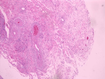
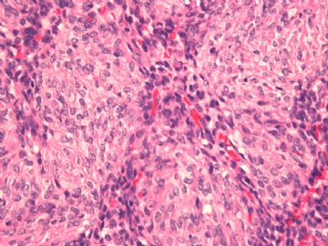
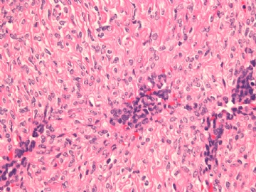
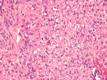
| 图片: | |
|---|---|
| 名称: | |
| 描述: | |
- 子宫透明细胞肿瘤2?
Adv Anat Pathol. 2008 Mar;15(2):63-75.
Perivascular epithelioid cell tumor (PEComa) of the uterus: an outcome-based clinicopathologic analysis of 41 reported cases.
Department of Pathology, Wilford Hall Medical Center, Lackland AFB, TX 78236, USA. oluwolefadare@yahoo.com
The uterus and retroperitoneum have emerged as the most frequently reported anatomic sites of origin of perivascular epithelioid cell tumors (PEComas), a poorly defined neoplasm that is characterized by varying amounts of spindle and epithelioid cells with clear to eosinophilic cytoplasm that display immunoreactivity for melanocytic markers, most frequently HMB-45. Published reports on 41 previously reported uterine PEComas are reviewed in this report. Of these 41 cases, 31 originating in the corpus and for which there was adequate follow-up information (or clinical malignancy) were categorized into 2 groups: (1) a malignant group that was comprised of cases associated with patient death of disease and/or clinical malignancy as evidenced by local and/or distant extension outside of the uterus (n=13, group 1) and (2) a "nonmalignant" group of cases in which neither of the above features were present (n=18, group 2). Groups 1 and 2 did not significantly differ regarding duration of follow-up (25 mo vs. 24.3 mo, respectively, P=0.9) or patient age (45.61 y vs. 43.46 y, respectively, P=0.7). Five of the group 1 patients experienced distant (extra-abdominal) metastases. The group 1 tumors were significantly larger than the group 2 tumors (averages 9.6 cm vs. 4.67 cm respectively, P=0.04); however, there were no size thresholds that, in of themselves, reliably classified 75% or more of the cases in both groups. Coagulative necrosis was highly associated with group 1, being present in 82% of cases as compared with only 11.8% of group 2 cases (P=0.0002). Eighty-eight percent of the group 2 cases had a mitotic rate of <or=1/10 high power fields (HPF) as compared with 40% of group 1 cases (P=0.01). However, the absence of mitotic activity did not rule out malignancy, as 2 of the group 1 cases lacked mitotic activity and displayed metastases. Twenty-five percent, 49%, 56%, 73%, and 100% of tested cases displayed immunoreactivity for CD10, desmin, vimentin, smooth muscle actin, and caldesmon, respectively. PEComas are tumors of uncertain histogenesis and malignant potential that seem to display some morphologic and immunophenotypic overlap with smooth muscle neoplasia. A mitotic count of >1/10 HPF and/or coagulative necrosis are features that, if present, raise the definite potential for aggressive behavior.

- 王军臣
Int Semin Surg Oncol. 2008 Mar 6;5:7.
Uterine PEComa: appraisal of a controversial and increasingly reported mesenchymal neoplasm.
Department of Pathology, Wilford Hall Medical Center, Lackland Air Force Base, San Antonio, TX 78236, USA. oluwolefadare@yahoo.com.
ABSTRACT: In recent years, a group of tumors that have been designated "perivascular epithelioid cell tumors" (PEComa) have been reported with increasing frequency from a wide variety of anatomic locations. The uterus and retroperitoneum appear to be the most frequent sites of origin for these lesions. PEComas belong to an identically named family of tumors comprised of conventional angiomyolipomas, clear cell sugar tumors, lymphangiomyomatosis and clear cell myomelanocytic tumor of the falciform ligament/ligament teres, and are also known as PEComa-NOS. This article is a primer for clinicians on the most salient clinicopathologic features of uterine PEComas, as most of the debate and discussion have taken place in the pathologic literature. The author appraises in detail the current state of knowledge on PEComas of the uterus based on a review of published data on the 44 previously reported cases, and comments on areas of controversy. The latter are centered predominantly on the significant morphologic and immunophenotypic overlap that exists between uterine PEComa and some smooth muscle tumors of the uterus. The clinicopathologic features of cases reported as epithelioid smooth muscle tumors and cases reported as uterine PEComas are compared and contrasted, and a practical approach to their reporting is proposed.

- 王军臣
-
需要鉴别恶性PEComa
Diagn Pathol. 2007 Dec 3;2:45.
Malignant perivascular epithelioid cell tumor (PEComa) of the uterus with late renal and pulmonary metastases: a case report with review of the literature.
Department of Pathology, University of Pittsburgh Medical Center, Pittsburgh, PA, USA. armahh2@upmc.edu.
ABSTRACT: BACKGROUND: Perivascular epithelioid cell tumor (PEComa), other than angiomyolipoma (AML), clear cell sugar tumor (CCST), and lymphangioleiomyomatosis (LAM), is a very rare mesenchymal tumor with an unpredictable natural history. The uterus is the most prevalent reported site of involvement of PEComa-not otherwise specified (PEComa-NOS). To the best of our knowledge, about 100 PEComa-NOS have been reported in the English Language medical literature, of which 38 were uterine PEComa-NOS. These reported cases of uterine PEComa-NOS have usually shown clinically benign behavior, but 13 tumors, three of them associated with tuberous sclerosis complex (TSC), exhibited local aggressive behavior and four of them showed distant metastases. CASE PRESENTATION: We report the case of a 59-year-old woman, who presented with renal and pulmonary lesions seven years after the initial diagnosis of uterine leiomyosarcoma. Left nephrectomy and right middle lobe wedge resection were performed. Histological and immunohistochemical analysis of the renal and pulmonary lesions, in addition to retrospective re-evaluation of the previous uterine tumor, led to the final diagnosis of malignant uterine PEComa with late renal and pulmonary metastases. All three lesions had the typical histological appearance of PEComa-NOS showing a biphasic growth pattern with continuous transition between spindle cells and epithelioid cells, often arranged around vascular spaces. Immunohistochemically, the tumor cells of both phenotypes in all three lesions stained for melanocytic (HMB-45 and Melan-A/MART-1) and myoid (desmin, smooth muscle actin, and muscle-specific actin/all muscle actin/HHF-35) markers. CONCLUSION: The findings indicate that despite the small number of reported cases, PEComas-NOS should be considered tumors of uncertain malignant potential, and metastases to other organs might become evident even several years after the primary diagnosis.

- 王军臣
-
需要注意有无性索分化的区域
Ann Diagn Pathol. 2010 Apr;14(2):129-32. Epub 2009 Aug 14.
A new morphological variant of uterine PEComas with sex-cord-like pattern and WT1 expression: more doubts about the existence of uterine PEComas.
Carvalho FM, Carvalho JP, Maluf FC, Bacchi CE.
Department of Pathology, Faculdade de Medicina da Universidade de São Paulo, São Paulo (SP), Brazil. filomena@usp.br
PEComas are rare neoplasms that are sometimes associated with the tuberous sclerosis complex. They typically contain perivascular epithelioid cells that coexpress muscle and melanocytic markers. However, apart from these classical features, considerable clinical, pathologic, and immunohistochemical variation has been reported. WT1, the Wilms tumor gene product, can be expressed in various tumors from different anatomical sites, including sex-cord and other ovarian tumors with a sertoliform pattern. Neither a sex-cord-like pattern nor WT1 expression has been described in PEComas. Here, we describe a case of uterine PEComa with a pattern of infiltration into the myometrium that is similar to stromal sarcomas, characterized by tongues and endovascular growing. The architecture and cellular morphology were similar to sex-cord tumors, and the PEComa was diffusely and strongly positive for WT1. We reviewed, from our files, an additional 9 cases of PEComa from different sites, and found WT1 expression in one more soft tissue tumor. We discuss the relationship between PEComas and other uterine sarcomas.

- 王军臣
| 以下是引用海上明月在2010-3-23 16:54:00的发言:
Int Semin Surg Oncol. 2008 Mar 6;5:7. Uterine PEComa: appraisal of a controversial and increasingly reported mesenchymal neoplasm.子宫PEComa:一个有争议的和逐渐报道的间叶肿瘤的评价 Department of Pathology, Wilford Hall Medical Center, Lackland Air Force Base, San Antonio, TX 78236, USA. oluwolefadare@yahoo.com.
ABSTRACT: In recent years, a group of tumors that have been designated "perivascular epithelioid cell tumors" (PEComa) have been reported with increasing frequency from a wide variety of anatomic locations. The uterus and retroperitoneum appear to be the most frequent sites of origin for these lesions. PEComas belong to an identically named family of tumors comprised of conventional angiomyolipomas, clear cell sugar tumors, lymphangiomyomatosis and clear cell myomelanocytic tumor of the falciform ligament/ligament teres, and are also known as PEComa-NOS. This article is a primer for clinicians on the most salient clinicopathologic features of uterine PEComas, as most of the debate and discussion have taken place in the pathologic literature. The author appraises in detail the current state of knowledge on PEComas of the uterus based on a review of published data on the 44 previously reported cases, and comments on areas of controversy. The latter are centered predominantly on the significant morphologic and immunophenotypic overlap that exists between uterine PEComa and some smooth muscle tumors of the uterus. The clinicopathologic features of cases reported as epithelioid smooth muscle tumors and cases reported as uterine PEComas are compared and contrasted, and a practical approach to their reporting is proposed. |
近年来,PEComa的报道逐渐增多,发病部位也较宽。子宫和腹膜后似乎是最常见的发病部位。PEComas属于血管平滑肌脂肪瘤同家族成员之一,透明细胞糖瘤,淋巴管肌瘤病和镰状韧带透明细胞肌黑色素细胞瘤/韧带,也是被称为PEComa-NOS。这篇文章是对子宫PEComas最突出的临床病理特征作了大部分讨论,已收在病理文献中。作者在先前报道的44例的基础上详细评价了子宫PEComas现状,并进行部分讨论。后者主要集中讨论了子宫PEComa和子宫平滑肌肿瘤的形态学和免疫组化上有些交叉重叠。对比和比较上皮样平滑肌瘤和子宫的PEComa的临床病理特征,提出切实可行的报告。

- 广州金域病理
| 以下是引用海上明月在2010-3-23 17:00:00的发言: 需要鉴别恶性PEComa Diagn Pathol. 2007 Dec 3;2:45. Malignant perivascular epithelioid cell tumor (PEComa) of the uterus with late renal and pulmonary metastases: a case report with review of the literature。子宫恶性PEComa伴后期肾脏和肺转移:1例报道及文献复习 Department of Pathology, University of Pittsburgh Medical Center, Pittsburgh, PA, USA. armahh2@upmc.edu. ABSTRACT: BACKGROUND: Perivascular epithelioid cell tumor (PEComa), other than angiomyolipoma (AML), clear cell sugar tumor (CCST), and lymphangioleiomyomatosis (LAM), is a very rare mesenchymal tumor with an unpredictable natural history. The uterus is the most prevalent reported site of involvement of PEComa-not otherwise specified (PEComa-NOS).To the best of our knowledge, about 100 PEComa-NOS have been reported in the English Language wymedical literature, of which 38 were uterine PEComa-NOS. These reported cases of uterine PEComa-NOS have usually shown clinically benign behavior, but 13 tumors, three of them associated with tuberous sclerosis complex (TSC), exhibited local aggressive behavior and four of them showed distant metastases.CASE PRESENTATION: We report the case of a 59-year-old woman, who presented with renal and pulmonary lesions seven years after the initial diagnosis of uterine leiomyosarcoma. Left nephrectomy and right middle lobe wedge resection were performed. Histological and immunohistochemical analysis of the renal and pulmonary lesions, in addition to retrospective re-evaluation of the previous uterine tumor, led to the final diagnosis of malignant uterine PEComa with late renal and pulmonary metastases. All three lesions had the typical histological appearance of PEComa-NOS showing a biphasic growth pattern with continuous transition between spindle cells and epithelioid cells, often arranged around vascular spaces. Immunohistochemically, the tumor cells of both phenotypes in all three lesions stained for melanocytic (HMB-45 and Melan-A/MART-1) and myoid (desmin, smooth muscle actin, and muscle-specific actin/all muscle actin/HHF-35) markers. CONCLUSION: The findings indicate that despite the small number of reported cases, PEComas-NOS should be considered tumors of uncertain malignant potential, and metastases to other organs might become evident even several years after the primary diagnosis. |

- 广州金域病理


