| 图片: | |
|---|---|
| 名称: | |
| 描述: | |
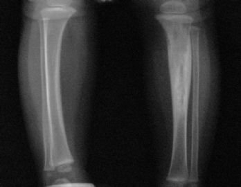
- 一岁男孩胫腓骨内病变,世所罕见(1991至今4例报道,最近到外地学习,又发现了一例,也是小朋友腿上)。
| 姓 名: | ××× | 性别: | 男孩 | 年龄: | 1岁 |
| 标本名称: | 胫骨活检 | ||||
| 简要病史: | |||||
| 肉眼检查: | |||||
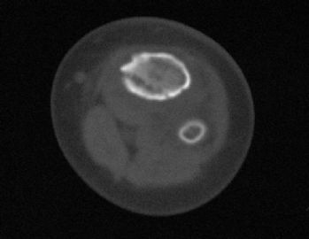
患儿,男,15个月,因被家人发现双下肢不等长并左小腿肿痛4月余,门诊拟诊“左胫骨中上段慢性骨髓炎”收入院。入院4月前,患儿开始学习走路,家人发现其站立不稳,双腿高低不平以及站立时患儿哭啼不适,逐到多家医院就诊,曾被疑为“脑瘫”的可能。外院X光片及CT片提示左胫、腓骨中上段骨病变,以胫骨病变明显,骨皮质不均匀增厚,骨干增粗,局部可见骨质增生及破坏性改变,拟诊“慢性骨髓炎”。入院时,患儿体重10.5Kg,发育正常,营养中等,神志清,精神好,查体合作。专科情况:双下肢不等长,左小腿较右侧长约2cm,局部肌肉无明显改变,中上段压痛,左踝、膝关节活动正常,趾动、血运及感觉正常。入院后在全麻下行左胫骨病灶活检手术。
-
本帖最后由 于 2010-09-12 22:13:00 编辑
-
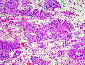
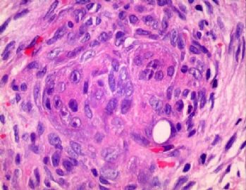
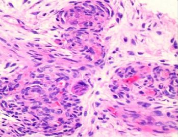
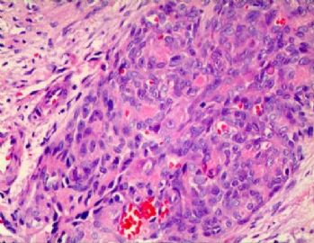
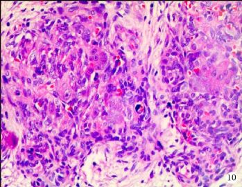
GCAB显著的特征是由卵圆形至梭形细胞构成的结节状、条素状和丛状结构。瘤细胞胞浆粉染,絮状;核呈圆形、肾形或梭形,核染色质呈弥散分布,有小核仁,偶见核分裂。
Common striking feature are nodular, linear, and plexiform aggregates of oval-to-spindle cells with pink flocculent cytoplasm and round, reniform, or spindle nuclei with dispersed chromatin, small nucleoli, and occasional mitoses
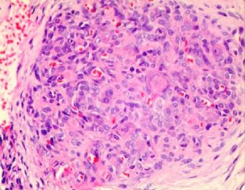
肿瘤结节内含有单核细胞和多核巨细胞(一个至多个核),常见明显核仁;胞浆丰富,含嗜酸颗粒。由卵圆形至梭形细胞以及巨细胞聚集而成的圆形结节,酷似非坏死性肉芽肿。
Within these aggregates were large mononucleate cells and multinucleate giant cells with one to several nuclei, often with prominent nucleoli, and abundant granular eosinophilic cytoplasm. Round conglomerates of oval-to-spindle cells and giant cells mimicked non-necrotizing granulomas.
有的结节中可见血管腔隙,中空或含有红细胞; 腔面衬覆胖圆形、立方形或扁平内皮细胞。这些小血管被卵圆形至梭形细胞围绕,呈同心性排列,具洋葱皮样表观。
Some aggregates had discernible lumens, either empty or containing erythrocytes, lined by plump, cuboidal, or flat endothelial cells.Concentric arrangement of oval-to-spindle cells around small-caliber vascular structures resulted in an onion skin appearance.
有的结节中,很少能见到包绕在病灶中的原有血管。偶见血管有内皮周细胞增生,呈偏心性生长,突向血管腔。邻近小血管处,易见肿瘤累犯神经膜或神经内。在有的区域,肿瘤结节之间含有疏松的间质,可见中等量的淋巴细胞和散在的肥大细胞浸润。
Envelopment of preexisting blood vessels rarely was observed.Occasional vessels showed eccentric periendothelial proliferation protruding into the lumen. Perineural and intraneural involvement by small vessels was common. In some regions, stroma between the tumoral aggregates was loose, and moderate lymphoplasmacytic infiltrate and scattered mast cells were present.
病灶周边血管增多,为毛细血管或稍大一些的血管,其内皮肥胖,可见一到两层内皮周细胞。间质疏松,含有纤细的小梭形细胞,血管分散其中。
Periphery of the lesions, increase in vessels of capillary or slightly larger size, with plump endothelial cells and generally one or two layers of periendothelial cells.Vessels were separated by a loose stroma containing delicate spindle cells.

- 王军臣
非常感谢Dr.Jin精确的诊断和天山望月提供详尽的文献资料。
在现版的WHO软组织分类中,归于其它的中间型血管肿瘤。
天山望月提供的资料大意如下:
巨细胞性血管母细胞瘤(Giant cell angioblastoma GCAB)是一种极其罕见的软组织肿瘤,为先天性肿瘤,或在幼婴时发生。迄今文献报道不足5例,发生的部位包括下唇、头皮和手部;均可形成溃疡,浸润周围组织甚至累及骨。 This rare soft tissue tumor and presents as a congenital tumor or in early infancy. Less than 5 cases have been described. These tumors have been described in the palate, scalp, and hand. All tumors were ulcerated and may infiltrate the surrounding soft tissue and bone.

- 王军臣
Background
This rare soft tissue tumor and presents as a congenital tumor or in early infancy. Less than 5 cases have been described. These tumors have been described in the palate, scalp, and hand. All tumors were ulcerated and may infiltrate the surrounding soft tissue and bone.
INCIDENCE Very rare AGE RANGE-MEDIAN Congenital or early infancy
HISTOLOGICAL TYPES CHARACTERIZATION General Common striking feature are nodular, linear, and plexiform aggregates of oval-to-spindle cells with pink flocculent cytoplasm and round, reniform, or spindle nuclei with dispersed chromatin, small nucleoli, and occasional mitoses
Within these aggregates were large mononucleate cells and multinucleate giant cells with one to several nuclei, often with prominent nucleoli, and abundant granular eosinophilic cytoplasm
Round conglomerates of oval-to-spindle cells and giant cells mimicked non-necrotizing granulomas
Some aggregates had discernible lumens, either empty or containing erythrocytes, lined by plump, cuboidal, or flat endothelial cells
Concentric arrangement of oval-to-spindle cells around small-caliber vascular structures resulted in an onion skin appearance
Envelopment of preexisting blood vessels rarely was observed
Occasional vessels showed eccentric periendothelial proliferation protruding into the lumen
Perineural and intraneural involvement by small vessels was common
In some regions, stroma between the tumoral aggregates was loose, and moderate lymphoplasmacytic infiltrate and scattered mast cells were present
Periphery of the lesions, increase in vessels of capillary or slightly larger size, with plump endothelial cells and generally one or two layers of periendothelial cells
Vessels were separated by a loose stroma containing delicate spindle cellsVARIANTS
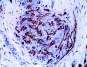
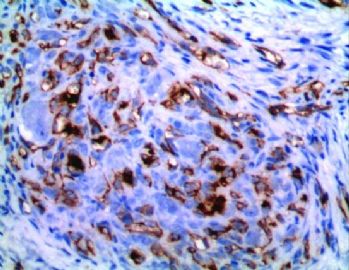
SPECIAL STAINS/IMMUNOPEROXIDASE/OTHER CHARACTERIZATION Special stains Immunoperoxidase Factor VIII and CD31 was positive in the endothelium of lesional and nonlesional vessels
Highlighted poorly discernible, small vascular channels within the densely cellular areas
Oval-to-spindle cells in the aggregates and periendothelial cells in the hemangioma-like areas showed strong positive staining for smooth muscle actin and vimentin
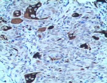
Strong positive staining for KP-1 antibody in the multinucleate giant cells and large mononucleate cells
Weakly positive staining for peanut lectin agglutinin in some of the multinucleate giant cells
Negative for:
Desmin, S-100, LCA, EMA, and Cam 5.2
DIFFERENTIAL DIAGNOSIS KEY DIFFERENTIATING FEATURES Giant cell fibroblastoma Express CD34 and show rearrangements of chromosomes 17 and 22 Epithelioid hemangioendothelioma with osteoclast-like giant cells Am J Clin Pathol 1990;93:813?.
Arch Pathol Lab Med 1993;117:315?.
Young adults
Epithelioid tumor cells in these cases, even in solid areas, recapitulated endothelial cells with intracytoplasmic lumina and Factor VIII stainingLack the concentric orientation of cells around small vessels
Plexiform fibrohistiocytic tumor Children and young adults and is characterized by the plexiform and nodular growth of rounded histiocytoid cells with interspersed osteoclast-like giant cell
Onion-skin layering of tumor cells around vessels and the hemangioma-like areas are absent
Myopericytoma Am J Surg Pathol 1998;22:513?5.
Cells with pericytic/myofibroblastic differentiation in a concentric arrangement around vascular lumens
Hemangiopericytoma-like areasGiant cells were not described in cases of myopericytoma
Found mainly in adults, youngest patient was 10 years old
Infantile myofibromatosis, infantile fibromatosis, or infantile fibrosarcoma Oncology Reports 1999;6:1101?.
Am J Surg Pathol 1994;18:14?4.
Characteristic chromosomal aberrations have been described
PROGNOSIS AND TREATMENT CHARACTERIZATION Prognostic Factors Natural history of giant cell angioblastoma is not fully known
Features typically associated with malignancy, such as a brisk mitotic rate, anaplasia, and necrosis are absent in giant cell angioblastoma
The clinical features, histological findings, and follow-up data suggest that giant cell angioblastoma is a benign tumor that would not be expected to metastasize but that could create significant local problems because of its infiltrative nature.
Treatment Am J Surg Pathol 1991;15:175?3.
This tumor extensively infiltrated the skin, soft tissue, and bone of the entire forearm-managed with amputation when the patient was 3 months old
Am J Surg Pathol 2001;25:185-196
With interferon- therapy one had no progression of residual microscopic disease
Second patient experienced considerable regression of the tumor with interferon- administered before subtotal excision-currently has stable minimal residual disease
Am J Surg Pathol 1991;15:175?3.
Am J Surg Pathol 2001;25:185-196

- 广州金域病理
-
X光片及CT片显示左侧胫骨、腓骨中上段骨病变,以胫骨改变明显,病变骨干明显增粗变形,髓腔内密度增高不均匀,骨皮质不规则增厚,骨内缘局部皮质缺损,邻近的软组织肿胀。镜下病变组织位于骨小梁之间,肿瘤组织呈结节状、粱索状或丛状聚集,瘤细胞卵圆形至梭形,散在胞浆丰富、核仁明显的单核组织细胞样巨细胞及多核巨细胞;肿瘤组织内可辨认出数量不等的无或含有少量红细胞的微血管腔隙;部分瘤细胞呈同心圆样围绕在微血管腔隙周围并与周围间质胶原共同形成洋葱皮样外观。在少数瘤结节的周边部,毛细血管扩张,充满红细胞或血清蛋白样物质,形态近似海绵状血管瘤。部分区域相邻瘤细胞排列成环状血管样腔隙,可有红细胞滞留;少数瘤细胞及巨细胞具有退变的趋势,未发现核分裂相、坏死及细胞的非典型性;肿瘤间质疏松,其间为少量梭形纤维母细胞样细胞、星形间质细胞、单核炎细胞及散在的肥大细胞。免疫组化标记显示卵圆形至梭形瘤细胞及巨细胞弥漫性强表达Vimentin,但一致性不表达Calponin、h-Caldesmon、EMA、AE1/AE3、CD1a;多量瘤细胞显著表达CD31及CD34,少数弱阳性表达FⅧ,环状血管样腔隙的瘤细胞亦可见前三者的表达;单核组织细胞样巨细胞及多核巨细胞强表达CD68,偶见个别巨细胞胞浆内局灶性表达FⅧ;肿瘤实质内小血管腔隙周围瘤细胞显著表达a-SMA,其他部分瘤细胞呈弱阳性表达;局灶性肿瘤组织内散在少量S100阳性的小梭形或多边形细胞,可能为朗格汉斯细胞。
显示详细信息 4:24 (6 小时前)
Certainly a very unusual and perhaps unique case. There are no more than 4 convincing examples of +++ in the English-speaking literature and the "entity" is quite controversial - but it seems that your case could fit. However, it is very difficult to make a certain diagnosis based on just a few selected images. Given the very unusual nature of this case, I would be glad to see glass slides of the case if you'd like to send materials to Boston (blocks would probably be best, in case we need to do more stains - obviously we will return the blocks promptly to you).
Kind regards
Chris Fletcher
