

| 图片: | |
|---|---|
| 名称: | |
| 描述: | |
- case1 宫颈小细胞癌; case2宫颈ADC; case 3 50 y/f月经增多
I showed a case here a few days ago. It was a very complicated law-suit case and I cannot find more photos now. So I deleted it because it is not good for education.Sorry for that.(几天前我贴了一个病例在这里。那是一个非常复杂的法律诉讼病例,由于我没有更多的图片,对于教学不是很好所以我删除了它。为此我深感抱歉。)
Our fellow showed an interesting case. I put here for your review.(我们的住院医有一个很有趣的病案,我贴在这里一起分享)
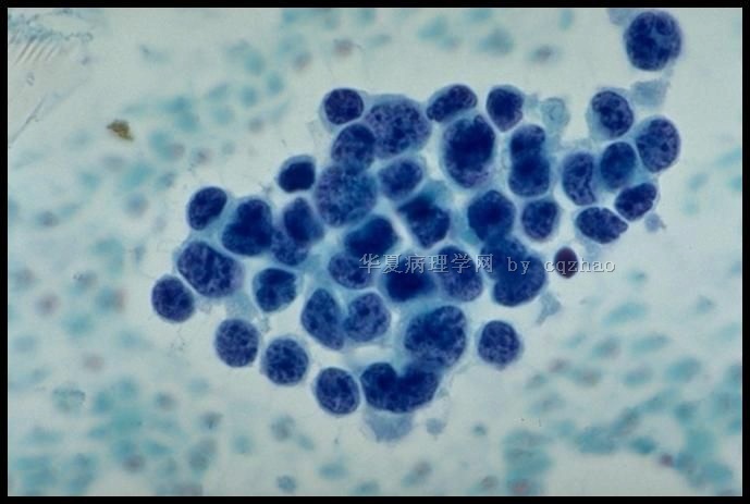
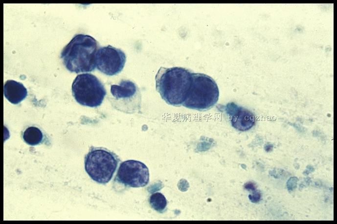
42 y women with LMP 5 days ago and no previous Pap history (女性,42岁,末次月经5天前,既往无巴氏检查)
标签:
-
本帖最后由 于 2010-05-07 07:28:00 编辑
×参考诊断
-
本帖最后由 于 2010-03-19 16:13:00 编辑
Benign endometrial cells
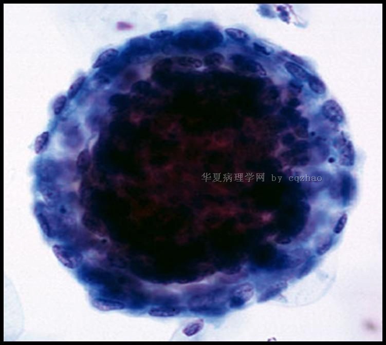
Condensed stromal cells surrounded by paler epithelial cells ("exodus balls")
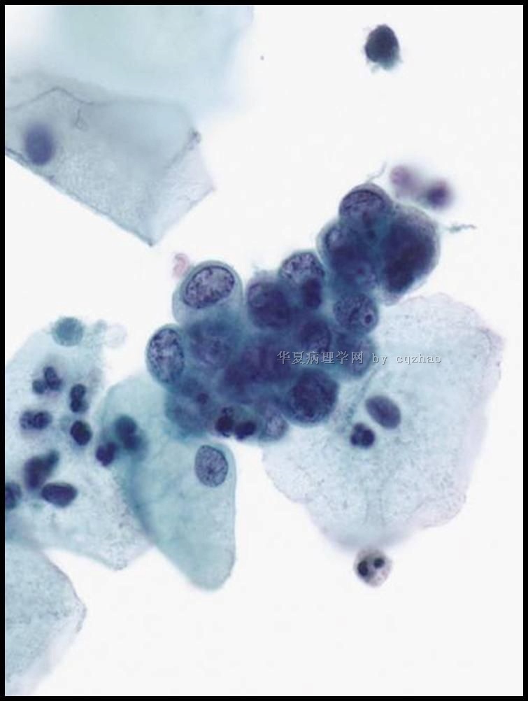
Closely packed epithelial cells w small hyperchromatic bean-shaped nuclei
Nuclear molding,variation in shape and size, apoptotic bodies
Cytoplasm scant, small vacuoles w engulfed PMN
1. Endometrial ball (exodus, present in LMP 6-10 days)
2. em cells
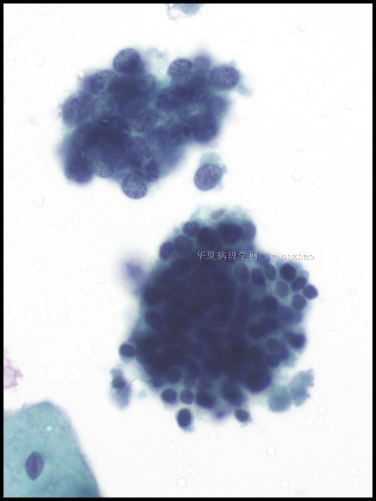
3. The case above: two clusters: upper small cell ca; low normal em cell
良性子宫内膜细胞
浓集的间质细胞周围围以栅状排列的上皮细胞(基质球)
密集成堆的上皮细胞,细胞小而深染,豆瓣形胞核,核扭曲,大小和形状多变,有时可见凋亡小体;胞质稀少,多形核细胞内可见卷入的小空泡
1、 子宫内膜基质球(LMP 6-10子宫内膜剥脱)
2、 子宫内膜细胞
3、 以上讨论本病例:两簇细胞,上面为小细胞癌,下面为正常子宫内膜细胞。
名称:图1
描述:图1
名称:图2
描述:图2
名称:图3
描述:图3
-
本帖最后由 于 2010-03-19 16:28:00 编辑
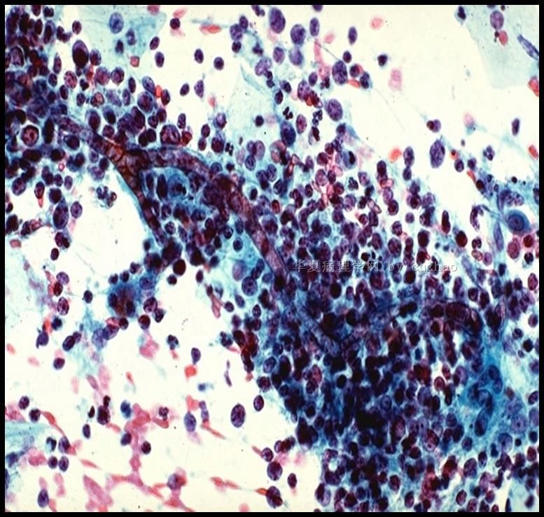
Follicular cervicitis
Lymphocytic cervicitis (lymphoid follicles in the subepithelial areas)
Cytology: mature and reactive lymphoid cells, macrophages w tingible bodies
~50% of the cases of follicular cervicitis associated with Chlamydia; also atrophy
Sometimes difficult to recognize in LBGS
– Small lymphoid cells in aggregates or dispersed
– Capillaries from the germinal center
– Should be distinguished for malignant lymphomas, histocytes, endometrial cells, and metastatic tumor cells
滤泡性宫颈炎
淋巴细胞性宫颈炎(上皮下存在淋巴滤泡)
细胞学:可见成熟的反应性淋巴细胞及巨噬细胞,约有50%衣原体相关的滤泡性炎可见可染小体(巨噬细胞吞噬淋巴细胞形成);
——聚集成堆或分散排列的小淋巴样细胞;
——有时可见生发中心来源的微血管;
——应该和恶性淋巴瘤、组织细胞、子宫内膜细胞和转移性肿瘤细胞相区分
名称:图1
描述:图1
| 以下是引用cqzhao在2010-3-13 16:33:00的发言:
When we read Pap, we should consider the patients, clinical situation, clinical managment, histology et al. These also can make the Pap more interesting. Some one can make a list for the differential diagnosis of small cell carcinoma in Pap test. Thanks, cz |
When we read Pap, we should consider the patients, clinical situation, clinical managment, histology et al.
These also can make the Pap more interesting.
Some one can make a list for the differential diagnosis of small cell carcinoma in Pap test. Thanks, cz
How do we tell difference of the small cell ca from different location? Some IHC stains may be helpful.
Histopathology. 2007 Sep;51(3):305-12.
Biomarker-assisted diagnosis of ovarian, cervical and pulmonary small cell carcinomas: the role of TTF-1, WT-1 and HPV analysis.
Carlson JW, Nucci MR, Brodsky J, Crum CP, Hirsch MS.
Department of Pathology, Brigham and Women's Hospital and Harvard Medical School, Boston, MA 02115, USA.
Comment in:
AIMS: Small cell carcinoma of the ovary, hypercalcaemic-type (SCCOH) is morphologically similar to small cell carcinomas from other sites. The aims of this study were to (i) determine if a biomarker panel would distinguish small cell carcinomas of the ovary, cervix (SCCCx) and lung (SCCLu) and (ii) potentially determine the histogenesis of SCCOH. METHODS AND RESULTS: Nine ovarian small cell carcinomas (seven hypercalcaemic type; two pulmonary type), eight SCCCx and 22 SCCLu were immunostained for thyroid transcription factor (TTF)-1, WT-1, p16, cKIT and OCT3/4; a subset of cases were tested for human papillomavirus (HPV). WT-1 was diffusely positive in 6/7 SSCOH versus two of 33 other small cell carcinomas (P <or= 0.001). TTF-1 was diffusely positive in 20/22 SCCLu and 1/8 SCCCx, and negative in all SCCOH. p16 and cKIT demonstrated variable patterns of immunoreactivity in all cases. HPV was identified in 5/6 SCCCx; SCCOH and SCCLu were negative for HPV. CONCLUSIONS: Combined staining with WT-1 and TTF-1 will distinguish SCCOH from SCCLu and SCCCx with a sensitivity of 86% and specificity of 97%. HPV is specific for tumours of cervical origin, but p16 immunohistochemistry is not useful for this purpose. The presence of diffuse WT-1 supports a Müllerian origin for SCCOH, whereas the absence of cKIT and OCT3/4 argues against a germ cell origin.
Immuno:
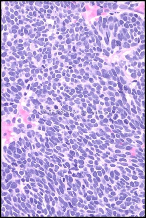
-Chromo+, Synapto+, CD56 (most senstitive) at least one neuroendocrine marker should be positive.
-p16+ almost all cases positive
CK (AE1/3)+, few case TTF-1 +,
Typical cytology of small cell ca
– Small cells (2xlymphs)
– Carrot-shaped nuclei
– Even powdery chromatin
– Nuclear molding
– Indistinct nucleoli
– Paranuclear blue bodies
– Mitoses
– Scant cytoplasm
– Background of nuclear debris and crush artifact (chromatin streaks)
-
本帖最后由 于 2010-03-13 16:07:00 编辑
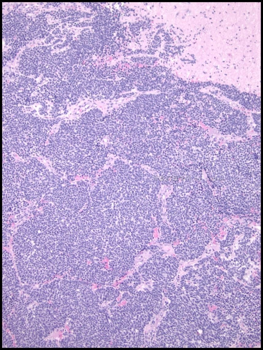
Some features of cervical small cell carcinoma
-Rare (2-5% of invasive cervical carcinomas)
-Clinically aggressive with rapid metastases; frequently presents with parametrial invasion and pelvic lymph node metastases
-Similar age as squamous cell carcinoma (mean 43 years, range 23 to 63 years)
-Associated with HPV, usually HPV-18
-Occasionally presents with paraneoplastic syndromes (Cushing etc)
-Coexisting SIL is rare; endocrine cell hyperplasia may be a precursor lesion
-Can be a part of squamous cell carcinoma or adenocarcinoma
| 以下是引用追逐太阳在2010-3-11 22:49:00的发言:
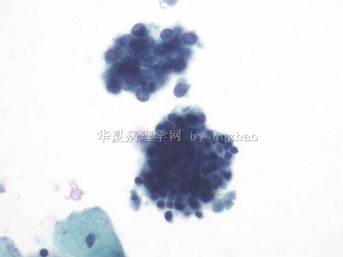 上面一团细胞,核:
1、小、圆,形态规则,大小一致,无差异性、多形性。
2、染色质呈细颗粒状,分布均匀,无集结、无块、无核仁。
3、纹理清晰,质感强,不是很透光,具有一定的立体感。
规则、一致+纹理清晰。这类细胞,如果出问题,问题会比较大,比如CIN3、类癌等。但鉴别诊断困难,即使考虑HSIL,也需要很大的细胞数量来作为支撑(相互验证、相互支持)。如果数量少、比例小,那问题也会很小,比如反应性改变,仅体现了一种增生活跃的状态。 |
Good analysis.
Are 上面一团细胞 and xia 面一团细胞 same or different? If low cluster is normal endometrial cells, 上面一团细胞 must be abnormal. This is called internal control.
小细胞神经内分泌癌占宫颈恶性肿瘤的1%到5%,并不是非常少见,但宫颈小细胞癌细胞学上均表现为中到高级别核型。肿瘤细胞常常单个散在或形成小簇,细胞小,圆形、卵圆形或不规则形,核深染,核仁不明显或缺乏,染色质细颗粒状,胞质稀少,核浆比高,有时可见核分裂相和核扭曲。这些特点本例不具备。
This is good.
But remember Text books always describe general situation. Every of your case in clinic may be unique.
Again Pap is a screening test.
We should not let this kind of case to have follow up only.
|
| 以下是引用掌心0164在2010-3-10 19:44:00的发言:
|
病人如果月经没有干净为什么要做检查?
Good quesion.
For Pap test, patient instruction:
1. better to have Pap sampling 2 week after the first day of the LMP
2. No vaginal medicine
3. No intercouse the night before the exam
My quesion is that Chinese patients or physicians always observe the rule for exam.


